A recent clinical trial with Organ-on-a-Chip devices has revealed a critical failure: fluid stagnation in microchannels caused massive cell death in the cultures. This incident underscores the need to validate microfluidic designs before manufacturing. The combination of medical segmentation software and computational fluid dynamics emerges as the most effective solution to predict these phenomena.
Technical workflow: From segmentation to fluid dynamics 🧪
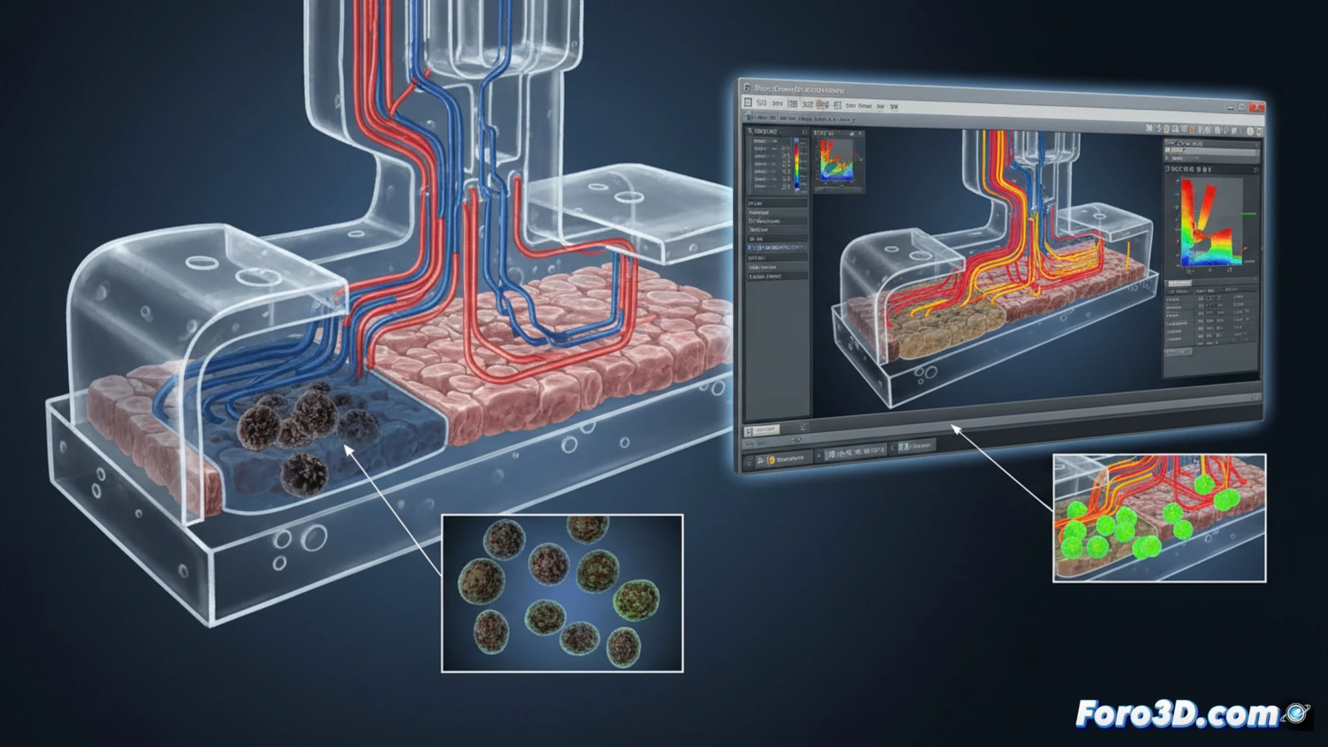
The process begins with Mimics, where CT scans or CAD designs are processed to generate a precise mesh of the chip geometry. This model is exported to Flow-3D, where the CFD solver is applied to simulate flow at the micrometer scale. In the analyzed case, Flow-3D detected recirculation zones and low velocity at the channel bifurcations, points where cell necrosis was later recorded. The simulation allowed visualizing pressure gradients and shear stress, revealing that the original design lacked diffusers to homogenize the flow rate. Blender was used to post-process particle trajectories and generate visualizations of the stagnant flow.
Lessons for biomedical chip design 🔬
This failure demonstrates that microfluidics cannot rely solely on geometric intuition. Integrating Flow-3D into the virtual prototyping phase allows identifying dead spots that compromise cell viability. For future trials, it is recommended to include parametric simulations that evaluate minimum flow rates and channel geometries with smooth transitions. Cell death not only invalidates the trial results but also delays drug development. Predicting these failures through CFD is now an indispensable requirement in tissue engineering.
Would you print this model in resin or filament? 🖨️