A postoperative leak after a robot-assisted intestinal resection set off alarms at a referral hospital. Initial suspicion pointed to surgeon error, but forensic analysis using micro-CT revealed a more complex truth: the titanium staples showed insufficient deformation. This finding shifted the investigation toward the robot's software and its ability to calculate the appropriate compression force based on tissue thickness.
Micro-CT reconstruction and finite element simulation in Abaqus 🧬
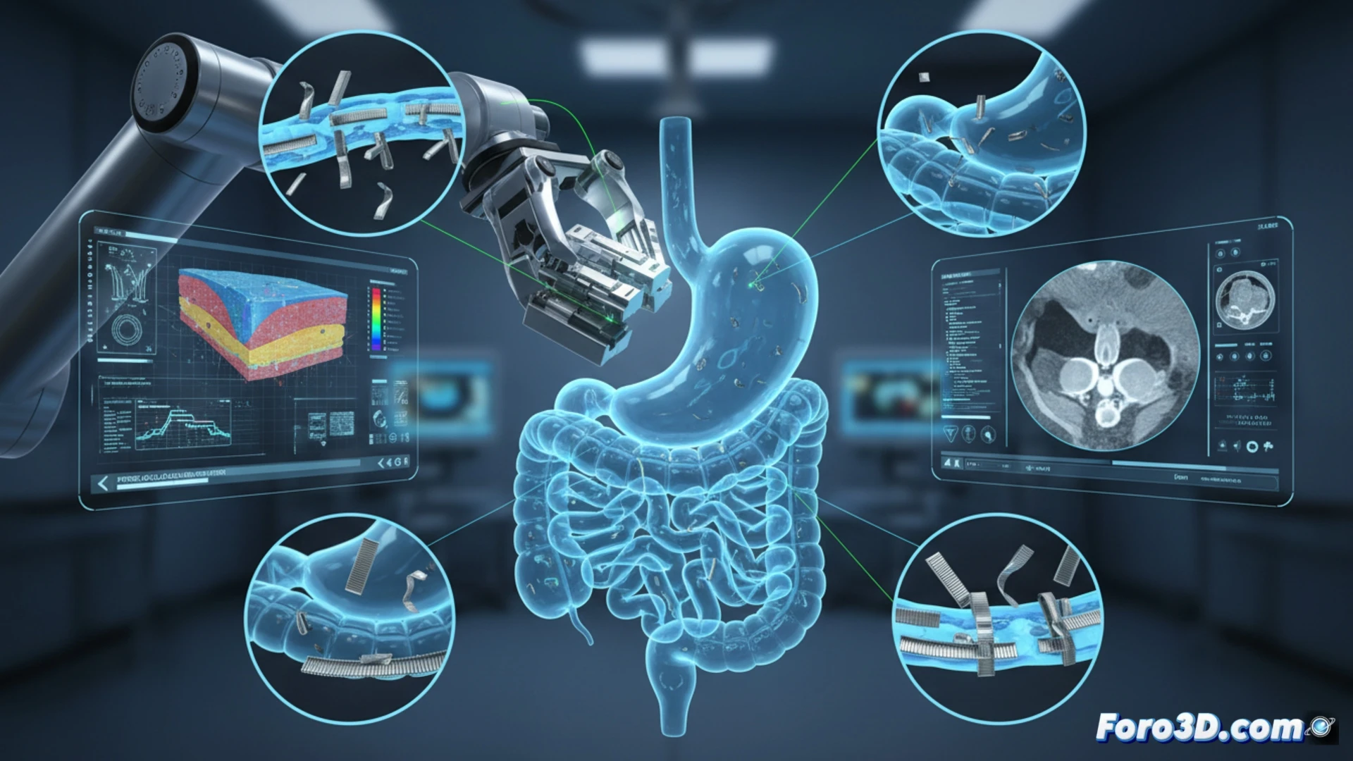
The forensic team digitized the extracted staples using micro-CT, generating high-resolution 3D models in Materialise Mimics. Comparing the actual geometry of the deformed staples with the ideal simulation in Abaqus revealed a critical discrepancy. The robot's software had applied a compression force equivalent to that for a 1.5 mm thick tissue, when the actual intestinal wall thickness was 2.8 mm. This underestimation caused the staples to fail to reach the closure height necessary to seal the tissue, leaving microchannels through which the leak occurred.
Robotic calibration: tissue thickness as a non-negotiable variable 🤖
The case demonstrates that the robot's mechanical precision is useless if the algorithms do not correctly integrate the patient's biomechanics. The lesson is clear: robotic surgery systems must calibrate their compression parameters in real-time, using sensors or preoperative CT data. Ignoring tissue thickness variability turns a high-tech tool into an avoidable risk for the patient.
How 3D analysis of the morphology and deformation of failed staples can be used to predict and prevent anastomotic leaks in gastrointestinal robotic surgery.
(PS: If you 3D print a heart, make sure it beats... or at least doesn't cause copyright issues.)