The Germans Trias i Pujol University Hospital has taken a firm step towards precision surgery. In less than two years, 205 interventions have benefited from personalized 3D models, created from real patient images. This technology allows surgeons to plan and rehearse each step before entering the operating room.
How a virtual twin of the patient is made 🏥
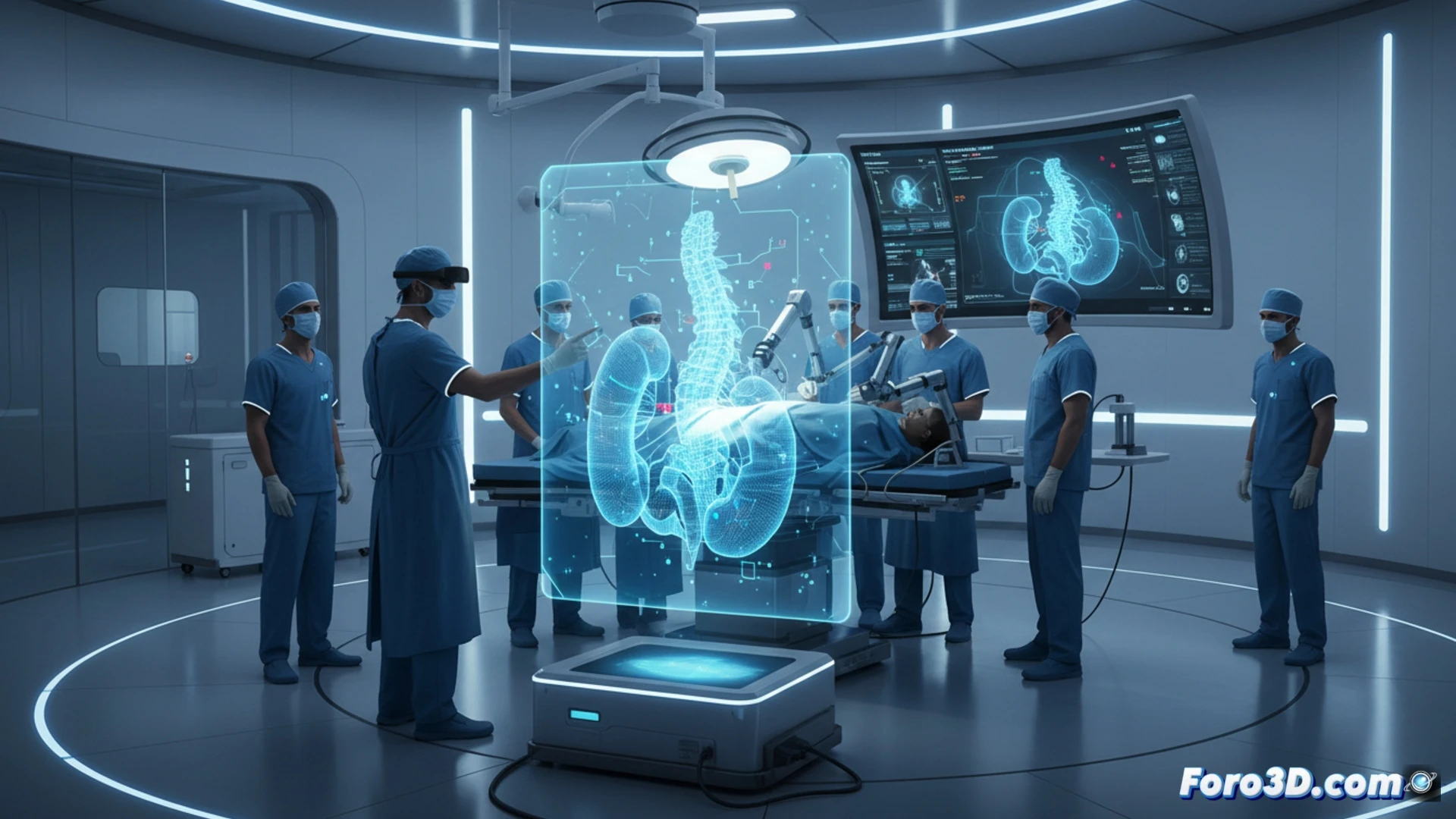
The process begins with a CT scan or MRI of the patient. This data is processed with specialized software to reconstruct the organ or structure to be operated on in 3D. The result is an exact model that is printed in plastic material or visualized in virtual reality. Surgeons use it to measure distances, simulate cuts, and anticipate complications, reducing risks and anesthesia time.
Goodbye to sketches on napkins 🛠️
Before, planning a complex surgery was like assembling Ikea furniture without instructions: a lot of faith and a few curse words. Now, with 3D models, surgeons can see the lump before making the incision. Of course, if the patient moves just before the print, the model might show a version of themselves that even their mother wouldn't recognize.