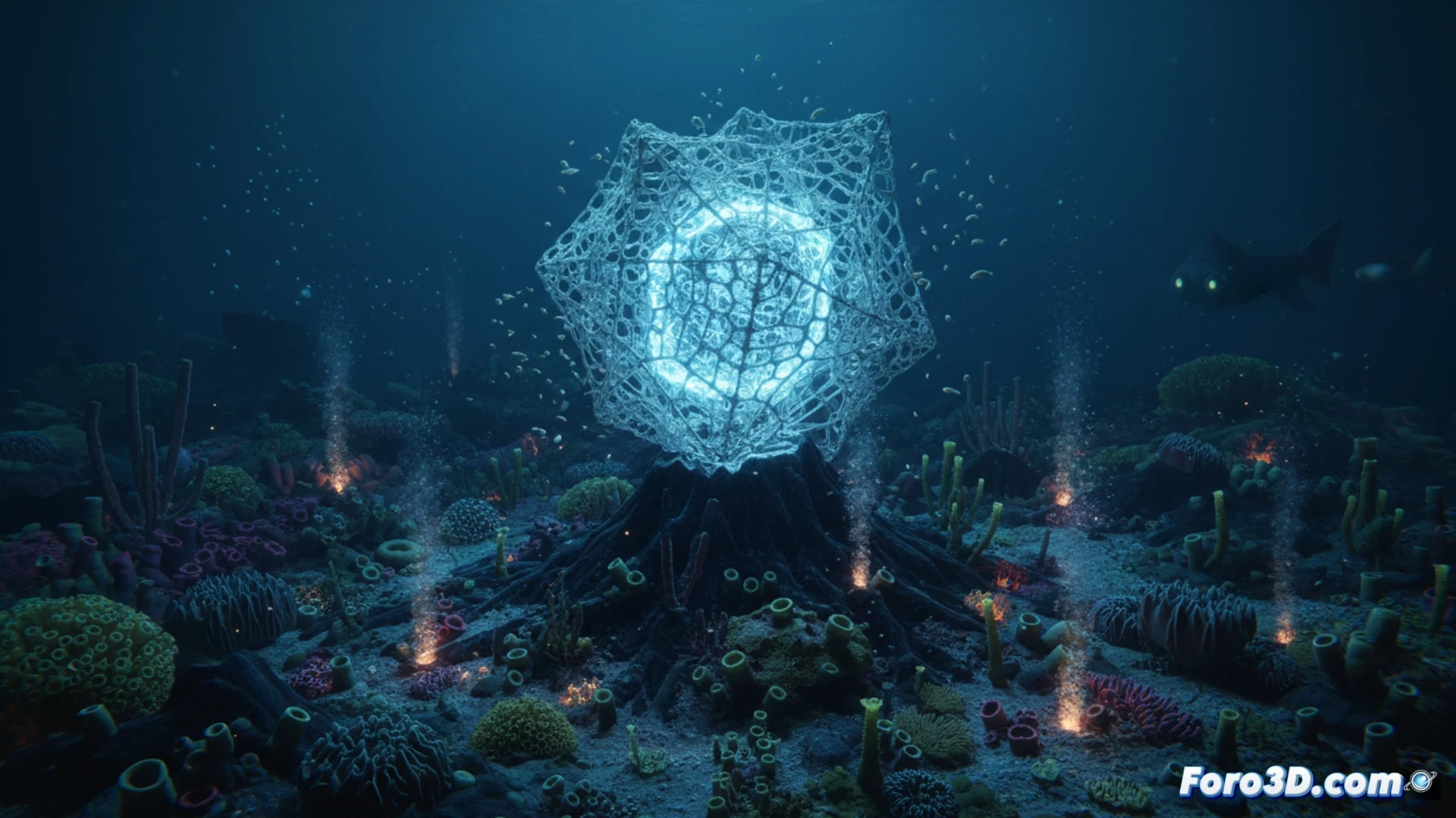
In 2024, the scientific community announced the discovery of a new species of glass sponge from the genus Walteria in the seamounts of Chile. What makes this organism exceptional is not only its biology, but its architecture: a skeleton of pure silica with a geometric complexity that challenges human engineering. For the niche of scientific visualization, this specimen represents a fascinating technical challenge, as its three-dimensional structure requires high-precision capture and rendering techniques to be fully understood.
Photogrammetry and microtomography for silica capture 🧬
The modeling of Walteria sp. cannot be done using traditional surface scanning methods, as its skeleton is composed of interwoven silica spicules that form an almost fractal three-dimensional grid. The most effective technique for digitizing this sponge is micro-computed tomography (micro-CT), which allows obtaining cross-sections of the specimen with micron resolution. From this data, a point cloud is generated, which is later reconstructed into a polygonal mesh. The result is a 3D model that can be visualized in software such as Blender or Houdini, where subsurface scattering shaders are applied to simulate the translucency of biological glass. This process allows marine biologists to rotate, section, and analyze the sponge without needing to dissect the actual specimen, thus preserving an extremely rare organism.
Aesthetics as a tool for scientific communication 🎨
Beyond pure research, the 3D visualization of Walteria sp. plays a crucial role in science communication. The beauty of its silica structure, reminiscent of a submerged Gothic cathedral, is a perfect visual hook to capture the attention of the general public. By generating high-resolution renders and orbital animations, scientists can explain complex concepts such as the biomechanics of poriferans or the biomineralization of silica in an intuitive way. The 3D model thus becomes a bridge between the laboratory and the audience, showing that nature remains the best designer of complex structures ever known.
Which 3D modeling tools and volumetric visualization techniques are most effective for representing the complex silica structure of the Walteria sp. glass sponge from computed tomography data?
(PS: at Foro3D we know that even manta rays have better social connections than our polygons)