In 2024, the herpetological community celebrated the discovery of the Brachycephalus lulai, a tiny toad barely one centimeter long, with a vibrant orange color, endemic to the cloud forests of Brazil. Named in honor of President Lula da Silva, this diminutive amphibian is not only a biological treasure but also a perfect candidate for advanced scientific visualization.
Precision Photogrammetry for a Cryptic Specimen 🐸
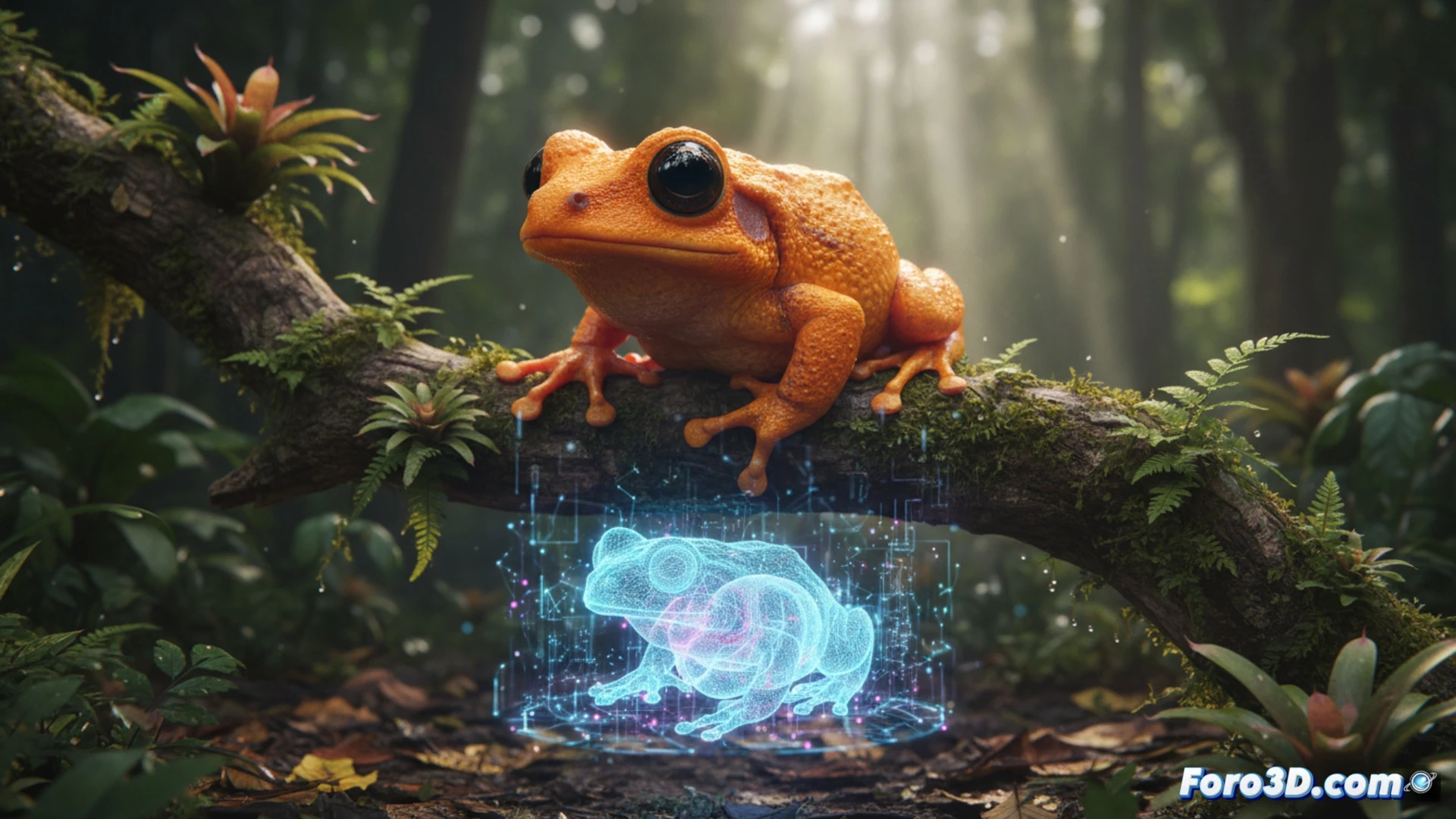
The fragility of Brachycephalus lulai, whose size and cutaneous toxicity make it difficult to handle, demands non-invasive documentation techniques. Through high-resolution photogrammetry, it is possible to capture every fold of its granular skin and the exact saturation of its aposematic pigmentation. The process involves taking hundreds of macro images with cross-polarized light to eliminate reflections, which are then processed in reconstruction software such as Agisoft Metashape. The result is a high-fidelity 3D mesh that allows researchers to measure the micro-ornamentation of the dermis without damaging the holotype.
The Cloud Forest as an Interactive Setting 🌿
Beyond the specimen, the ecological context is vital. Digitally reconstructing the understory of the Serra do Mar, with its layer of moist leaf litter and constant mist, offers a virtual laboratory to study the camouflage and behavior of this tiny toad. An animation showing it jumping among bromeliads, rendered with global illumination in Unreal Engine, allows educators to explain its evolution without the need for costly expeditions. Thus, 3D scanning becomes a conservation tool, democratizing access to a species that, due to its rarity, few could observe in the wild.
What specific technical challenges did researchers face when digitally scanning a specimen only one centimeter long, like Brachycephalus lulai, and how did they solve problems such as capturing microscopic details in its anatomy without damaging the living fossil?
(PS: fluid physics for simulating the ocean is like the sea: unpredictable and you always run out of RAM)