The rupture of a heart valve in companion or farm animals is not only a clinical emergency but also a challenge for biomechanical understanding. 3D technologies, from CT-based modeling to replica printing, now allow us to isolate the structural failure and visualize the deformation of valvular tissue. This technical analysis explores how these tools are redefining the study of heart failure in the veterinary field.
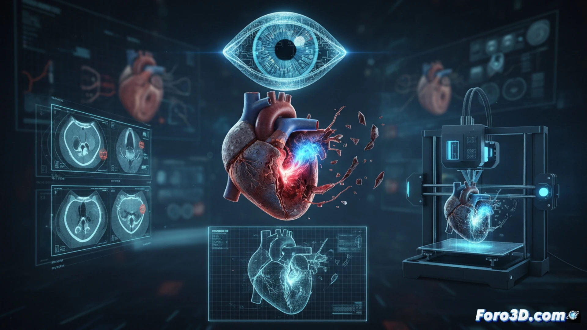
Modeling and simulation of structural valvular failure 🩺
Three-dimensional reconstruction of the animal heart from DICOM images allows for the creation of a digital twin of the affected valve. Using finite element software, researchers can simulate the mechanical stresses that precipitate rupture, identifying fatigue zones in the valvular collagen. These simulations, validated with 3D printing in flexible materials such as TPU, offer a tactile replica for planning reconstructive surgeries. In cases of endocarditis or myxomatous degeneration in dogs, the printed model guides the surgeon in suturing or prosthetic replacement, reducing ischemia time and improving implant precision.
Custom prostheses and the future of veterinary cardiology 🐾
Valvular rupture reveals the need for prostheses that mimic the native geometry of the animal. 3D printing allows for the design of custom biocompatible valves, adjusted to the diameter of the valvular annulus and the patient's flow dynamics. This approach not only saves lives but also turns each structural failure into a lesson in tissue engineering. The anatomical replica thus becomes the central tool for understanding why a valve fails and how we can reconstruct it with millimeter fidelity.
How 3D modeling of animal valvular structure allows predicting biomechanical failure points before they occur in real tissue.
(PS: If you 3D print a heart, make sure it beats... or at least doesn't cause copyright issues.)