Precise documentation of sharp-force injuries is a cornerstone of criminal investigation. 3D technology, through photogrammetry and laser scanning, allows capturing the exact geometry of a wound, overcoming the limitations of traditional photography. This approach digitizes evidence for objective and reproducible analysis, essential in the modern forensic pipeline.
Technical workflow: injury capture and modeling 🛠️
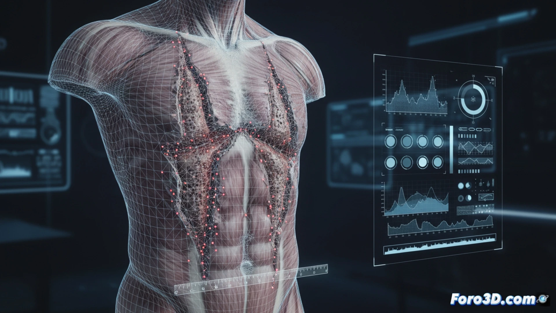
The process begins with data acquisition. For external wounds, high-resolution photogrammetry is used, taking multiple images with cross-polarized light to eliminate shadows and capture tissue texture. For deep wounds, it is combined with handheld laser scanning to obtain the three-dimensional morphology of the entry channel. Modeling software (such as MeshLab or CloudCompare) aligns the point clouds, generating a precise mesh. On this mesh, penetration vectors and attack angles are calculated, simulating the weapon's trajectory. Integration with 3D models of the suspected weapon allows comparing the blade width and wound pattern, validating or refuting expert hypotheses.
Digital objectivity as the new evidentiary standard ⚖️
The transition from visual inspection to 3D simulation eliminates subjective biases in interpreting wound depth or angle. An expert report based on a digital model allows the jury to visualize the dynamics of the attack without relying on static photographs or ambiguous verbal descriptions. This method, although technically demanding, raises the standard of forensic evidence by converting a biological injury into quantifiable and verifiable data in three-dimensional space.
What technical limitations does photogrammetry present for accurately reconstructing the trajectory and depth of a stab wound in postmortem human tissue?
(PS: In the forensic pipeline, the most important thing is not to mix the evidence with the reference models... or you'll end up with a ghost at the scene.)