The use of hydrogels as intervertebral implants promises natural integration, but their mechanical strength remains a critical point. A recent forensic analysis has managed to unravel the causes of a fracture in an explant of this material. Through a workflow combining high-precision scanning and finite element simulation, the exact moment of failure has been reconstructed. This case demonstrates how 3D biomechanics becomes an indispensable tool for the validation of medical devices. 🔬
Forensic workflow: From segmentation to simulation 🛠️
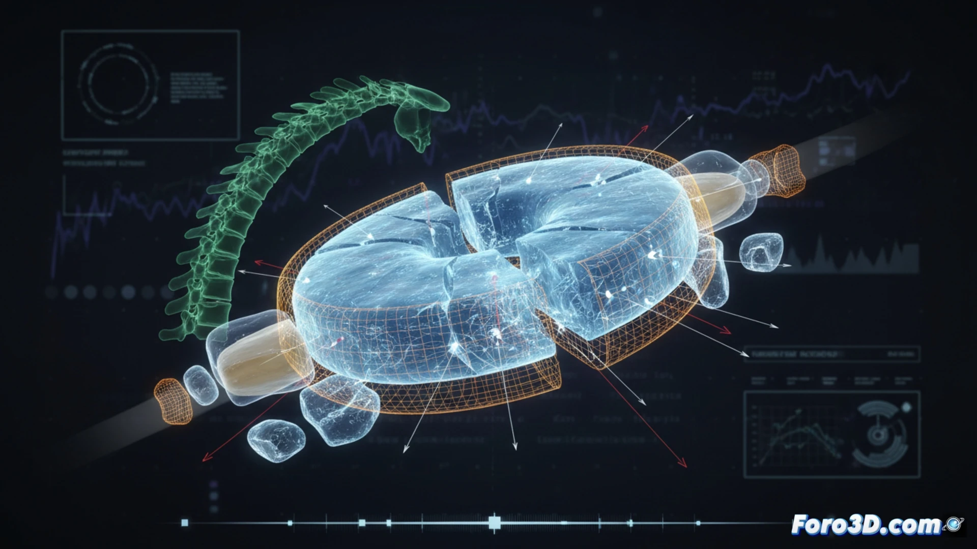
The process began with the digitization of the fractured explant. Using Mimics, the geometry of the damaged implant was segmented, separating the base material from the fracture lines to obtain an accurate solid model. This model was exported to Ansys, where typical physiological loading conditions of the lumbar segment were applied. The finite element simulation revealed stress concentration in the fracture area, identifying a shear fatigue point. Finally, 3ds Max allowed for the creation of a failure visualization, overlaying stress maps onto the actual explant geometry to communicate the results clearly.
Lessons for designing safer implants 💡
The analysis not only explains the failure but also provides a roadmap for improving the design. The simulation showed that the original implant geometry generated an excessive stress point at the interface with the vertebral bone. Thanks to the 3D reconstruction, engineers can now modify the internal architecture of the hydrogel to distribute loads more evenly. This predictive approach, combining scanning and simulation, is key to avoiding future surgical revisions and increasing the lifespan of biomedical implants.
How 3D modeling the progression of mechanical failure in an intervertebral hydrogel helps predict its durability before clinical trials
(PS: If you 3D print a heart, make sure it beats... or at least that it doesn't cause copyright issues.)