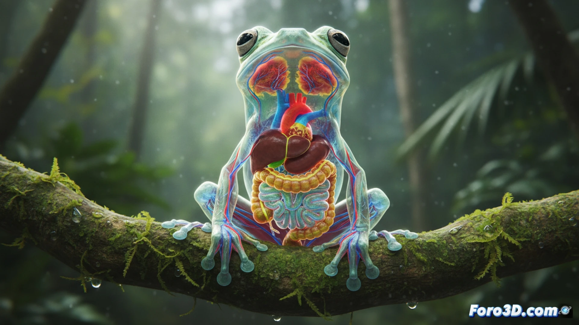
The discovery of the Hyalinobatrachium dianae in Costa Rica has captured the attention of biologists and artists alike. Its transparent ventral skin allows for live observation of the heart, liver, and digestive tract, a characteristic that makes it ideal for scientific visualization. This article explores how to create an interactive 3D model that not only reproduces its anatomy but also serves as an educational tool to explain biological transparency and evolution. 🐸
Photorealistic modeling and transparency system 🔬
To represent the frog's transparency in a 3D engine, two material layers must be configured: one for the dorsal skin (opaque and green) and another for the ventral skin (translucent with a refractive index of 1.33). Internal organs, such as the heart and bones, must be modeled with independent geometry and assigned a subsurface scattering (SSS) material type to simulate light dispersion. The environment should recreate the Costa Rican understory with a moisture particle system and HDRI-based tropical rainforest lighting. The animation can include a breathing cycle that expands the thoracic cavity, making the heart's movement visible through the skin.
Visual comparison and reflection on outreach 🎭
The accidental similarity to the Kermit the Frog puppet invites us to reflect on how nature surpasses fiction. By including a comparative scene where the 3D model of the real frog is placed alongside a stylized model of the puppet (with its characteristic neck and bulging eyes), we can highlight the real anatomical differences. This contrast is not only a visual hook but also allows explaining concepts such as biological mimicry versus artistic design, demonstrating that 3D models can be bridges between science and popular culture.
What 3D modeling techniques allow accurately representing the transparency and internal anatomy of the Kermit glass frog for scientific outreach?
(PS: at Foro3D we know that even manta rays have better social connections than our polygons)