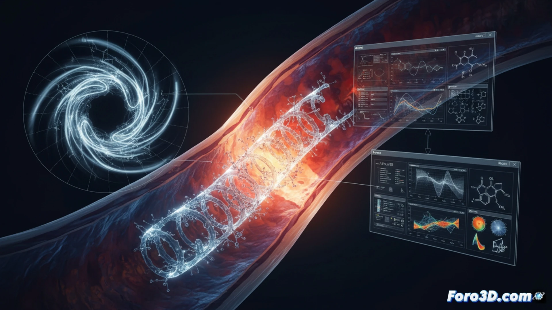
Early re-stenosis in polymer coronary stents has challenged the reliability of these devices. 3D forensic analysis, supported by OCT (Optical Coherence Tomography), allows mapping the material degradation rate to determine if hydrolysis occurred faster than designed. This technical article breaks down the multidisciplinary workflow combining MATLAB, Abaqus, and Materialise Mimics to model and predict the premature failure of these critical implants.
Workflow: From OCT to Finite Element Model 🔬
The process begins with the segmentation of OCT images in Materialise Mimics, where the 3D geometry of the stent is reconstructed and mass loss due to hydrolysis is identified. This point cloud is exported to MATLAB for detailed volumetric analysis, calculating the localized degradation rate along the stent struts. Subsequently, the generated mesh is transferred to Abaqus, where cyclic loading conditions typical of the coronary environment are applied. The material fatigue simulation, fed with real hydrolysis data, reveals stress concentration zones that accelerate polymer fracture before the expected time.
The Importance of 3D Forensic Analysis in Implant Certification 🛡️
The convergence of these tools allows going beyond a simple failure analysis. A direct correlation is established between the degraded microstructure and the mechanical response of the stent. 3D forensic analysis not only determines the cause of re-stenosis but also provides critical data to redefine polymer design parameters. In a sector where patient life depends on model precision, material fatigue simulation consolidates itself as the ultimate forensic tool to validate the safety of implantable medical devices.
How can 3D fatigue simulation predict the exact fracture point due to hydrolysis in a bioresorbable stent before early re-stenosis occurs?
(PS: Material fatigue is like yours after 10 hours of simulation.)