Accurate assessment of a muscle strain is critical for athletic performance. Thanks to computational biomechanics and 3D modeling, specialists can now visualize damage to muscle fibers with millimeter precision. This approach allows for the creation of digital twins of the athlete to simulate tension in the injured tissue, optimizing diagnosis and reducing the risk of relapses during competition.
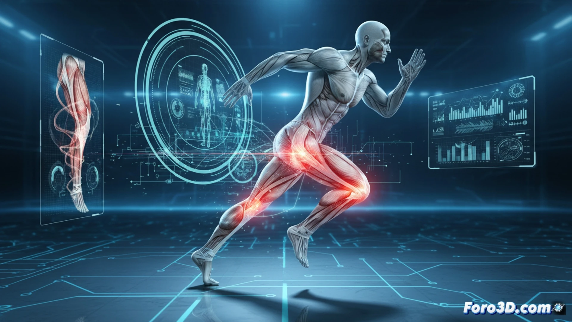
Biomechanical simulation and volumetric reconstruction of damaged tissue 🏋️
The process begins with data capture using magnetic resonance imaging and 3D surface scanners, which are integrated into finite element simulation software. The three-dimensional model of the muscle allows for the application of vector forces that replicate specific sport movements, such as a sprint or a change of direction. By visualizing the stress distribution in the strain area, physiotherapists can predict the injury's progression and adjust training loads. This technology transforms subjective assessment into a quantitative analysis of the tissue's structural integrity.
The future of predictive rehabilitation in sports 🚀
The implementation of muscle digital twins not only accelerates recovery but also redefines injury prevention. By simulating fatigue or overload scenarios, coaches can modify an athlete's technique before a rupture occurs. This convergence between 3D technology and sports medicine marks a turning point in athlete health management, shifting from a reactive model to a predictive and personalized one.
How can 3D modeling based on computational biomechanics predict the exact recovery time of a muscle strain in elite athletes?
(PS: player tracking is like following your cat around the house: lots of information and little control)