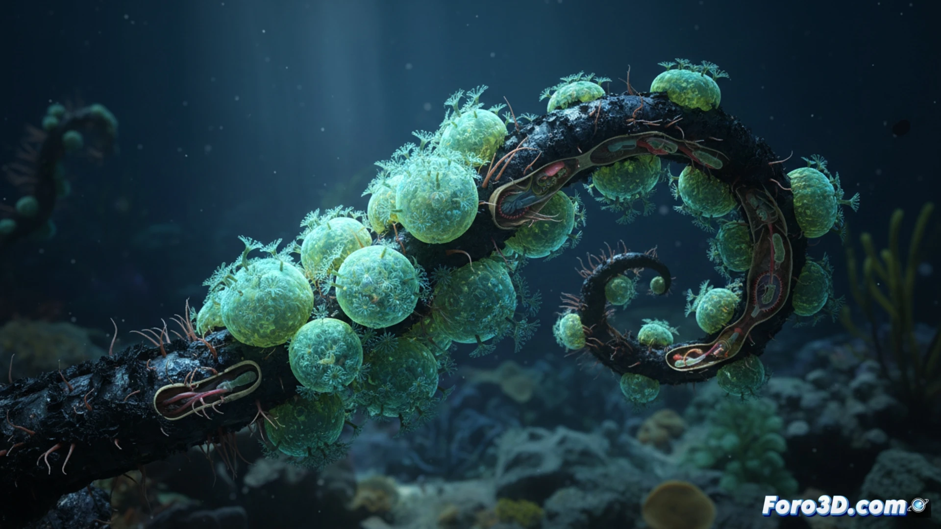
A recent biological-marine discovery has documented a unique symbiotic relationship: a zoanthid, similar to a sea anemone, that grows exclusively on the skeletons of certain black corals in the depths of Nazca. This finding, which challenges the traditional understanding of benthic interactions, requires scientific visualization tools to be communicated effectively. Three-dimensional representation emerges as the ideal vehicle to dissect the parasite's morphology and its attachment to the host.
Technical Workflow: From Underwater Photogrammetry to Symbiosis Animation 🐙
The modeling process begins with field data capture. Using underwater photogrammetry with high-resolution cameras and controlled lighting structures, a point cloud of the colonized black coral is obtained. Concurrently, scanning electron microscopy (SEM) provides cross-sections of the zoanthid polyp, revealing the attachment zone and tissue penetration into the calcareous skeleton. In Blender or ZBrush, the coral mesh is reconstructed from the point cloud, while the zoanthid is digitally sculpted using SEM references. The final animation shows a time-lapse of the parasite's growth, highlighting the spatial relationship through cross-sections that illustrate symbiosis at the cellular level. The model is exported in OBJ and GLTF formats for publication in repositories such as Sketchfab and MorphoSource.
The Value of Visualization in Deep Ecology Outreach 🌊
Beyond scientific rigor, this 3D model serves as a bridge between the laboratory and the general public. By allowing any user to digitally explore a deep-sea ecosystem, access to a finding that would otherwise be invisible is democratized. The animation, by dynamically showing the parasitic interaction, transforms an abstract concept into a striking visual experience. Publishing this work on Foro3D not only documents the technical process but also invites the community to reflect on how technology can make tangible the fragility and complexity of life in ocean trenches.
What are the main technical challenges when 3D modeling the morphology of a parasitic zoanthid and its symbiotic interaction with black coral, considering the lack of detailed anatomical data from this recent discovery?
(PS: modeling manta rays is easy, the hard part is making them not look like floating plastic bags)