Shaken infant trauma, known as shaken baby syndrome, represents one of the most devastating injuries in pediatrics. The biomechanics of brain damage are complex to analyze in vivo. This is where biomedical 3D modeling becomes an essential tool. Through segmentation of magnetic resonance imaging (MRI) and computed tomography (CT) images, specialists can virtually reconstruct the infant's skull and brain to study the impact of acceleration and deceleration forces.
Segmentation and Rendering of Diffuse Axonal Injury 🧠
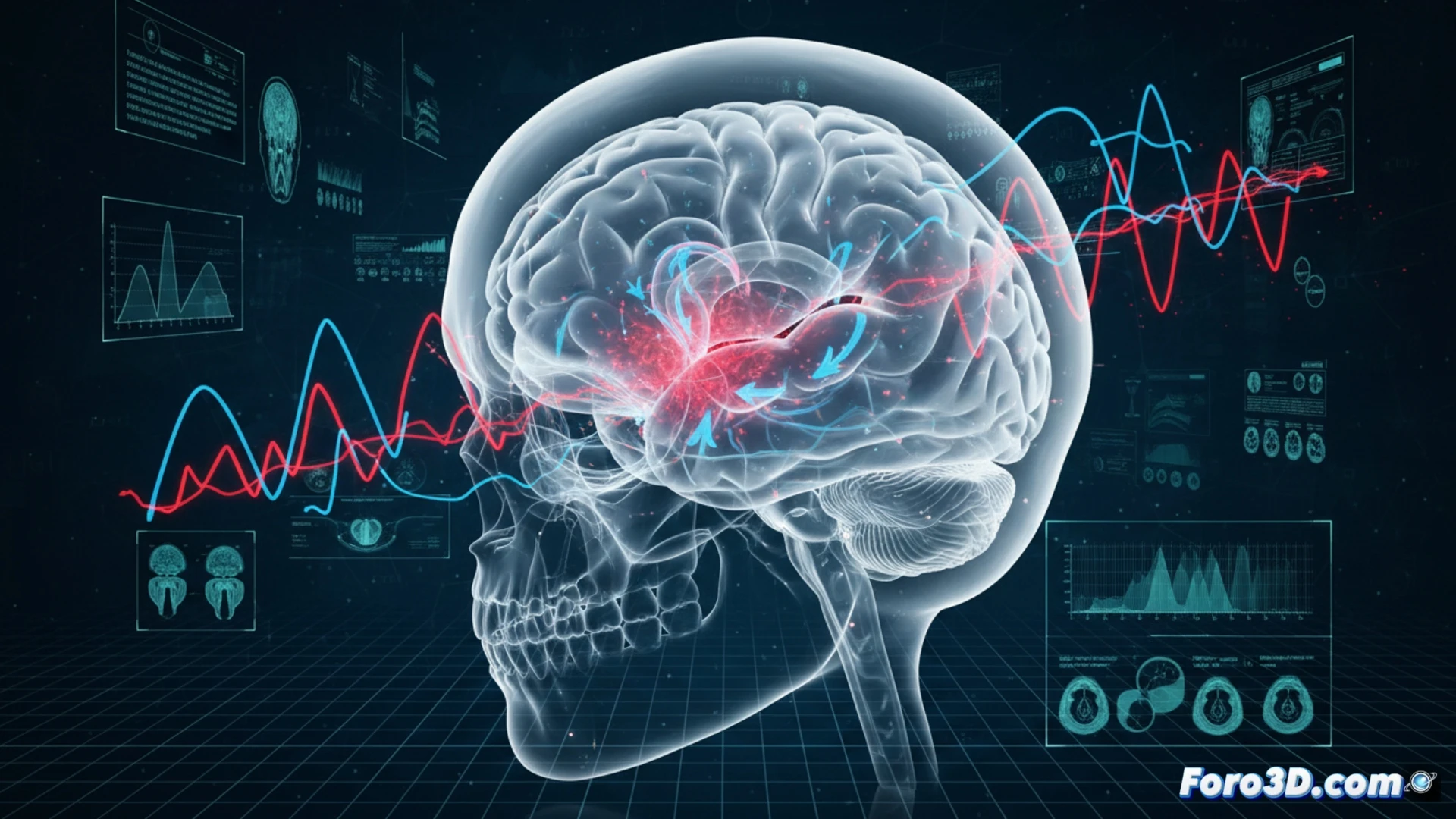
The technical process begins with the acquisition of high-resolution DICOM data. Using software such as 3D Slicer or Mimics, semi-automatic segmentation of intracranial structures is performed. The goal is to isolate subdural hemorrhages and cerebral edema, key characteristics of abuse. Subsequently, volume rendering algorithms are applied to visualize the distribution of injuries. These 3D models allow forensic experts to simulate the injury mechanism, calculating force vectors and differentiating between accidental trauma and violent shaking. The precision of the polygonal mesh is critical to avoid losing details of fissures or blood vessels.
The Ethics of Visualizing Invisible Damage ⚖️
Beyond technique, these models serve a social and judicial function. By converting abstract medical data into tangible three-dimensional representations, communication of the damage to judges or juries is facilitated. However, the modeler must be rigorous: a misinterpretation of a hemorrhage in the render could bias a diagnosis. The technical writer's responsibility is to document each step of the segmentation, ensuring that the 3D model serves as objective scientific evidence and not as a speculative recreation of violence.
How can biomechanical 3D modeling help differentiate injuries from shaken infant trauma from those caused by accidental falls in forensic practice?
(PS: and if the printed organ doesn't beat, you can always add a little motor... just kidding!)