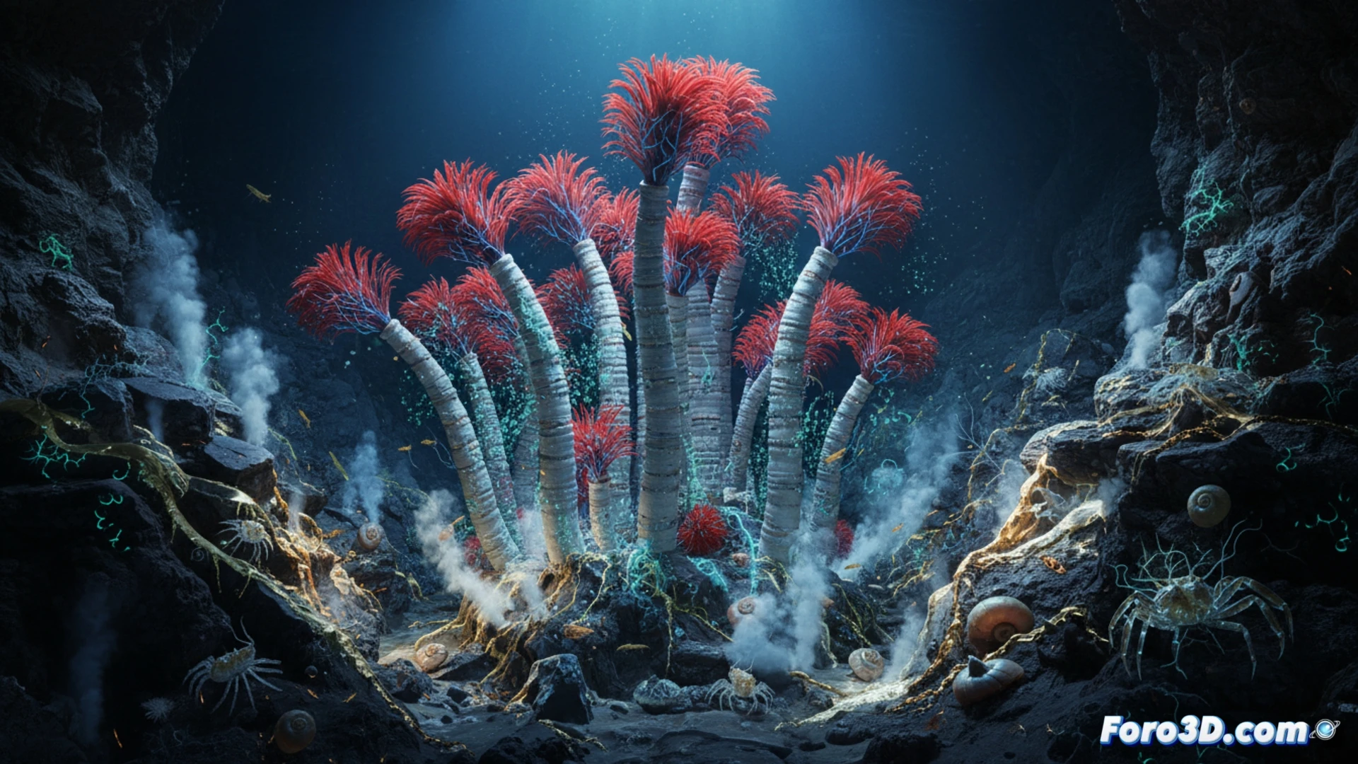
The sulfur tubeworm (Escarpia sp.) presents a fascinating challenge for 3D scientific visualization. This organism, which inhabits cold seep environments, builds extensive gardens of calcareous tubes and relies on internal chemosynthetic bacteria to metabolize hydrogen sulfide. Modeling its anatomy, bacterial symbiosis, and the flow of minerals in its environment allows researchers to simulate extreme ecosystems, offering clues about life on other planets and in the deep ocean. 🐛
Anatomical Modeling Techniques and Flow Simulation 🌀
To recreate Escarpia sp. in 3D, it is recommended to start with a base model of the worm using NURBS curves to capture its vermiform body and characteristic red branchial plume. The protective tube should be modeled separately, applying a procedural displacement with calcium carbonate textures. The real technical challenge lies in simulating the vascular system and the trophosome chamber where the symbiotic bacteria reside. Here, volume shaders and particle systems are ideal for visualizing the exchange of compounds such as sulfide and methane. Additionally, the laminar flow of cold fluids around the tubes must be simulated, using real-time or pre-calculated fluid simulations to show how dissolved minerals reach the worm. The lighting should be dim and bluish, replicating abyssal conditions, with volumetric light points to simulate the faint chemiluminescence of the habitat.
Implications for Astrobiology and Science Communication 🌌
Visualizing Escarpia sp. is not just an exercise in biological realism; it is a tool for conceptual exploration. By modeling this ecosystem, scientists can generate hypotheses about what life might be like on icy moons such as Enceladus or Europa, where hydrothermal vents or cold seeps exist. An interactive model, where the user can dissect the worm and see the flow of energy from mineral to bacteria and then to the animal, transforms an abstract concept of chemosynthesis into a tangible experience, vital for education and space mission planning.
What 3D modeling techniques allow for the most accurate representation of the transparency and bioluminescence of the Escarpia tubeworm's tissues in a chemosynthetic deep-sea environment?
(PS: modeling manta rays is easy; the hard part is making them not look like floating plastic bags)