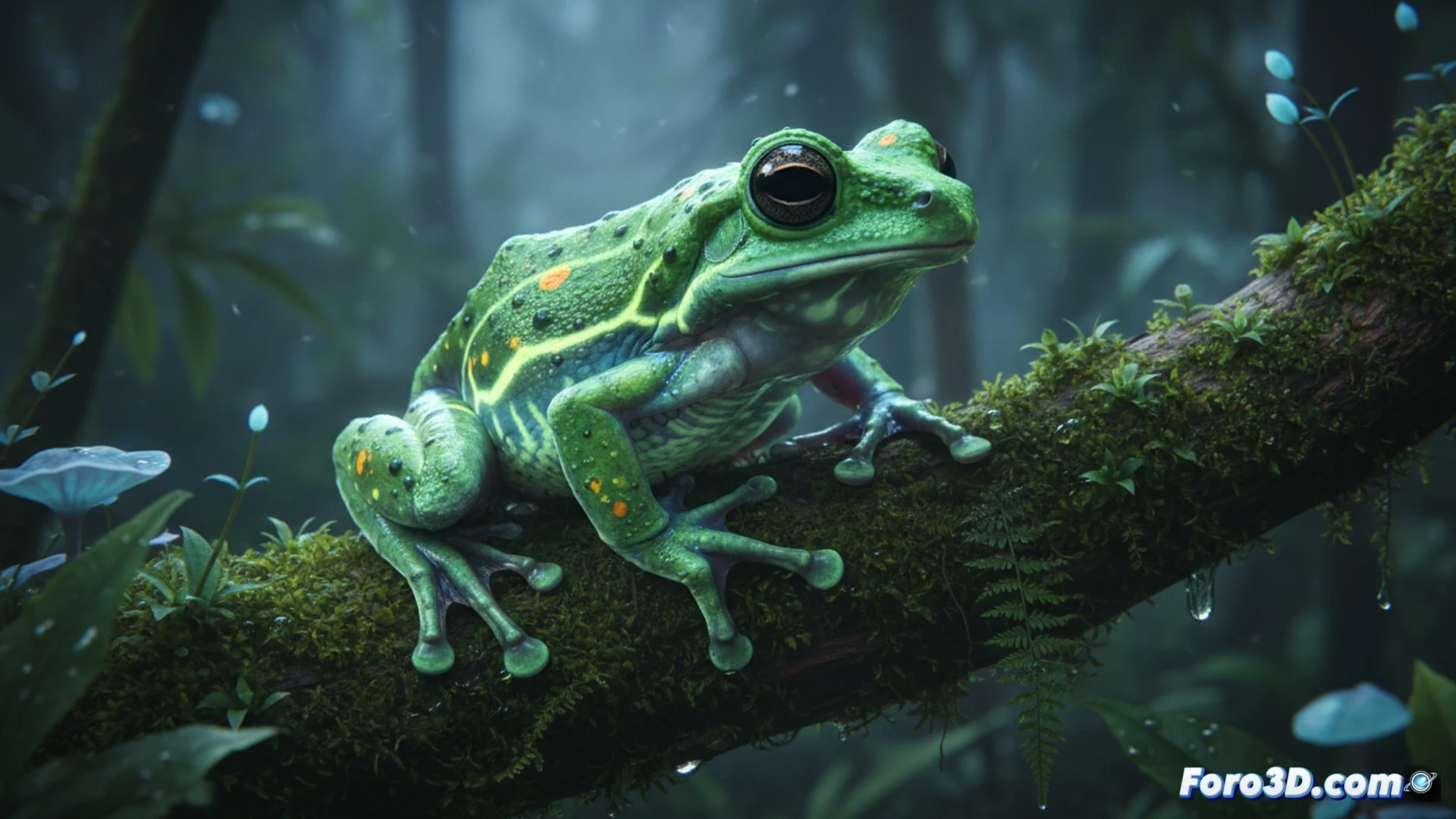
The discovery of Hyloscirtus jaramilloi in the cloud forests of Ecuador represents a milestone for modern herpetology. Its emerald green skin speckled with purple dots is not only an aesthetic marvel but also a technical challenge for scientific visualization. This article explores how photorealistic 3D modeling can capture the bioluminescence and texture of this species, facilitating its study without the need for physical capture.
Technical Reconstruction of Habitat and Anatomy 🐸
To faithfully represent Hyloscirtus jaramilloi, it is crucial to develop a high-resolution polygonal model that reproduces the microstructure of its glandular skin. The key lies in displacement mapping to simulate the purple speckles and a shader based on subsurface scattering (SSS) that emulates the translucent emerald tone. Additionally, recreating the Andean environment requires particle systems for perpetual mist and epiphytic vegetation, along with volumetric lighting that simulates light filtered through the forest canopy. This allows researchers to analyze the frog's camouflage in real time.
Visualized Genetics and Immersive Outreach 🧬
The greatest challenge of this project is translating genetic data into an interactive infographic. Using procedural nodes, we can link specific DNA markers to variations in the color and morphology of the 3D model, visually showing the differences between Hyloscirtus jaramilloi and its close relatives. This scientific animation not only validates the taxonomic study but also brings the complexity of evolution closer to a general audience, transforming a journal article into an immersive experience for conservation.
Considering the challenges of translucency and skin microstructure of Hyloscirtus jaramilloi, what shading and 3D texture mapping techniques would allow for more accurately replicating its emerald green coloration and the optical properties of its epidermis for use in scientific visualization?
(PS: at Foro3D we know that even manta rays have better social connections than our polygons)