The Nepenthes wallacei, discovered in Indonesia and named in honor of Alfred Russel Wallace, represents a fascinating challenge for scientific 3D modeling. Its purple pitchers, capable of retaining large insects, demand an accurate representation of their functional anatomy. This technical article explores the process of creating a detailed digital asset, from the peristome topology to the simulation of digestive fluid, aimed at educational documentaries and interactive museum exhibits.
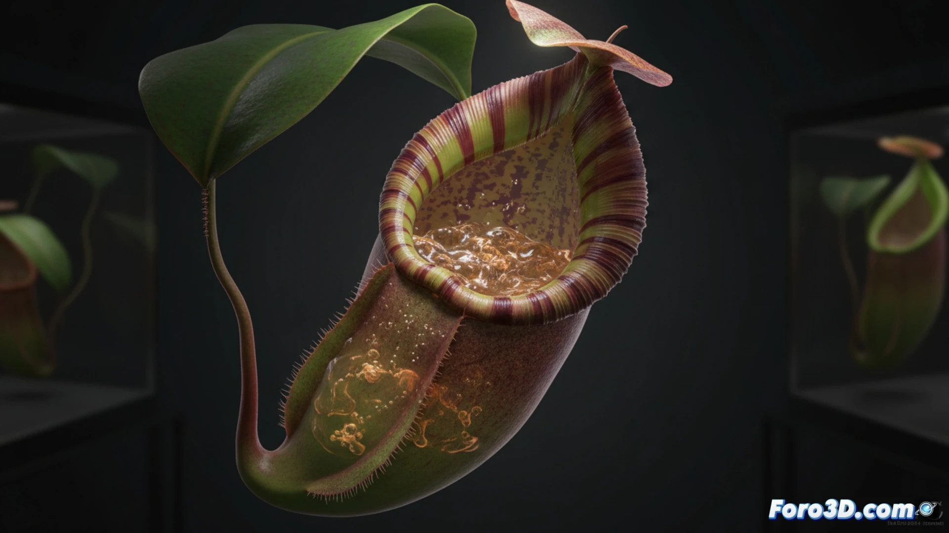
Digital anatomy and simulation of the capture mechanism 🧬
For the base model, it is recommended to use photogrammetry of preserved specimens combined with LiDAR scanning of the habitat in Sulawesi. The pitcher geometry must include a ribbed peristome with high polygon density to represent the marginal teeth that secrete nectar. The PBR texture of the operculum requires a roughness map that simulates the epicuticular wax, a key factor in the slippery inner surface. For the simulation of the capture process, a particle system is implemented that emulates the insect's movement towards the digestive fluid, using a soft body physics engine. The animation of the lid closure must be synchronized with the detection of insect collisions against the inner wall, a crucial detail for scientific accuracy in visualization.
The value of precision in science communication 🔬
Beyond technical realism, this 3D model allows biologists and educators to demonstrate the convergent evolution of Nepenthes. By including a visual comparison with Dionaea muscipula (Venus flytrap) and Drosera, understanding of different carnivory strategies is facilitated. The interactive simulation of the digestive fluid pH, visualized through a color gradient inside the pitcher, turns an abstract concept into a tangible experience. For a virtual museum, this asset not only documents a species, but reconstructs its ecological niche, making Indonesia's biodiversity accessible to any user.
How can 3D modeling of Nepenthes wallacei reveal hidden biomechanical patterns in its structure to improve the accuracy of ecological and evolutionary simulations in scientific visualization?
(PS: modeling manta rays is easy, the hard part is making them not look like floating plastic bags)