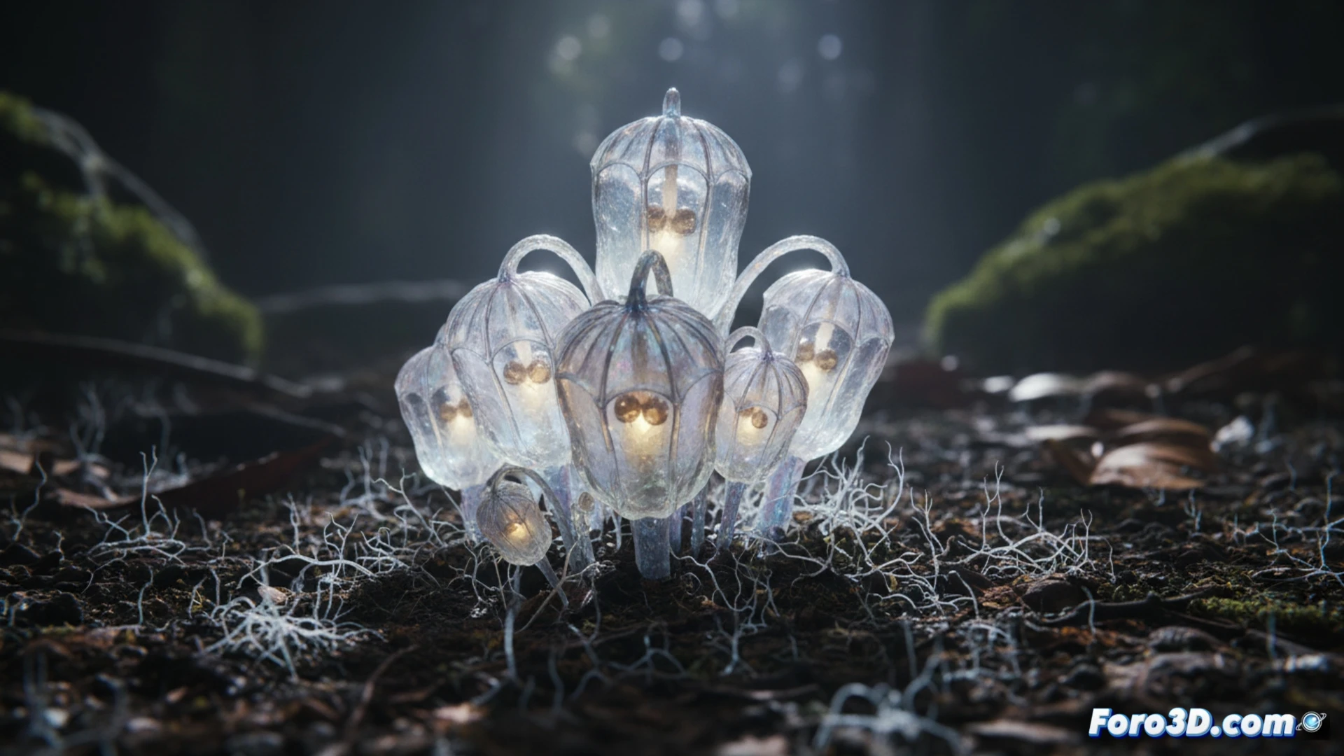
The Thismia selangorensis, known as the Selangor Fairy Lantern, is a mycoheterotrophic plant that defies the rules of botany. Discovered in the tropical rainforests of Malaysia, this species does not perform photosynthesis; instead, it emerges from the soil solely to bloom, displaying a translucent structure resembling a glowing lantern. Its unique biology makes it a perfect candidate for scientific visualization, where 3D modeling can capture its ephemeral morphology and symbiotic relationship with underground fungi.
Modeling Techniques for Translucent Structures 🌿
To photorealistically recreate Thismia selangorensis, priority must be given to simulating its translucency and lantern-like shape. Polygonal modeling should begin with a high-resolution photogrammetric scan of a preserved specimen, followed by retopology to optimize the mesh. Texturing requires subsurface scattering (SSS) maps that mimic light filtering through its tissues, with a pale orange color and semi-transparent veins. The emergence animation from the soil is achieved through particle simulations for the substrate and soft rigging that deforms the stem as the plant rises. The cross-section should include a network of fungal hyphae modeled with splines and volumetric shading to represent the mycorrhiza. Scientific data such as the absence of chlorophyll and dependence on fungal nutrients will be integrated into interactive annotations within the 3D viewer.
The Poetics of the Invisible in Visualization ✨
Modeling the Fairy Lantern is not just a technical exercise; it is an act of making the invisible visible. This plant reminds us that life in the jungle does not only occur above ground, but in an underground network of chemical exchanges. By creating a 3D model of its emergence and its cross-section, we are translating an almost alien biological process into a comprehensible visual language. The final animation should evoke the fragility of its existence, illuminating how beauty can arise from darkness and total dependence on another organism.
What specific technical challenges does the three-dimensional reconstruction of Thismia selangorensis present, considering its translucent structure and underground mycorrhizal symbiosis, and how can they be overcome to achieve accurate scientific visualization?
(PS: modeling manta rays is easy; the hard part is making them not look like floating plastic bags)