The discovery of the carnivorous sponge Abyssocladia johnhooperi in 2024 has opened a new frontier for scientific visualization. Its most distinctive feature is the hook-shaped spicules it uses to capture prey on the seafloor. Named in honor of poriferan expert John Hooper, this species represents a fascinating technical challenge for 3D modeling, where each microstructure must be accurately represented to understand its hunting mechanism.
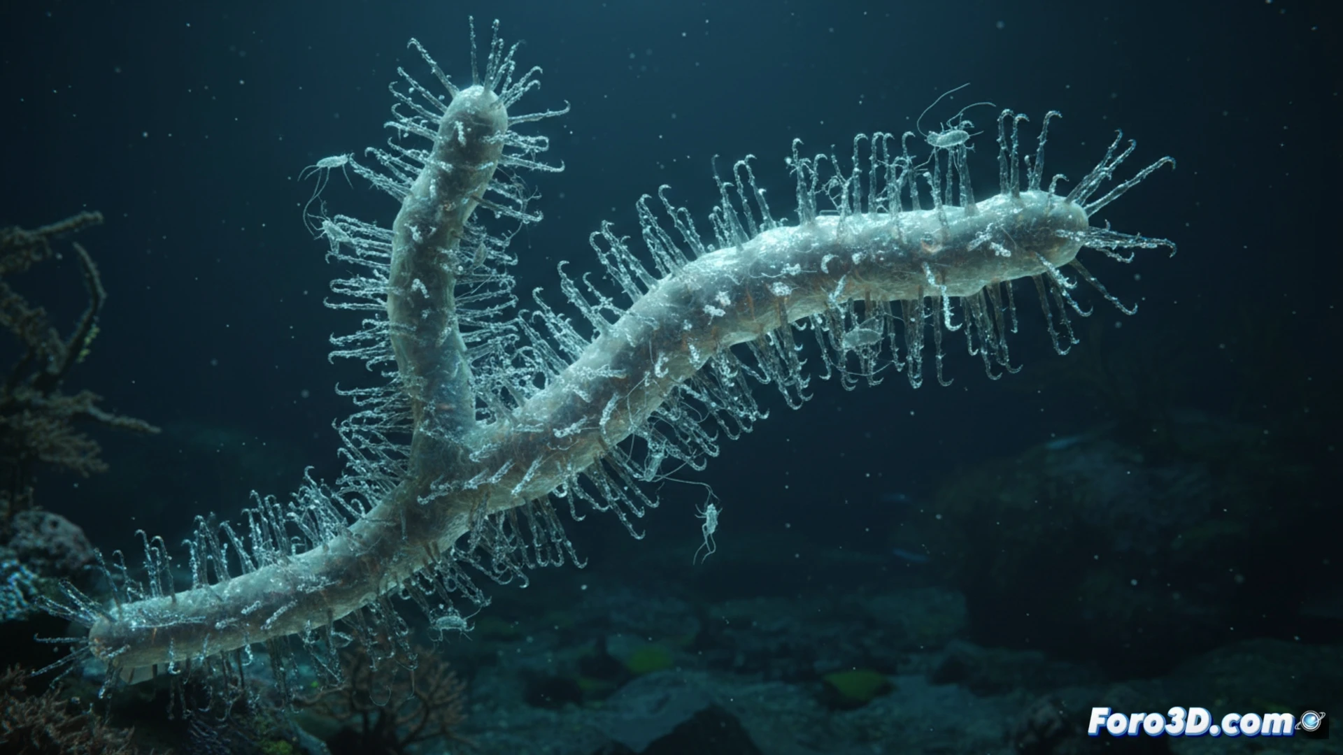
Microscopy Rendering and Capture Animation 🧬
For an accurate representation, the 3D model must integrate scanning electron microscopy data. The spicules, composed of silica, require texture mapping that simulates their transparency and refraction. In Blender or Maya, a particle system can be used to recreate the canopy of adhesive filaments surrounding the sponge. Animating the capture mechanism involves simulating the contraction of these filaments, similar to a lasso, trapping small crustaceans. A workflow using photogrammetric scanning of real specimens would enhance the model's authenticity for use in scientific publications and documentaries.
The Potential of Visualization in Marine Biology 🌊
The digital recreation of Abyssocladia johnhooperi not only beautifies an article but serves a crucial educational function. By comparing its spicule architecture with that of other sponges in the genus Abyssocladia, biologists can study the evolution of carnivory in deep-sea environments. An interactive model allows rotating and zooming in on the sponge, revealing details that a two-dimensional photograph hides. This approach turns a laboratory discovery into an immersive visual experience, ideal for virtual museums and marine biology classrooms.
How can the complex structure of hooks and spicules of Abyssocladia johnhooperi be modeled in 3D to accurately represent its prey capture mechanism in scientific visualization?
(PS: modeling manta rays is easy, the hard part is making them not look like floating plastic bags)