The discovery of the genus Mini in Madagascar has revolutionized herpetology. With species such as Mini mum, Mini scule, and Mini ature, these frogs do not exceed one centimeter in length. For a scientific visualization editor, this finding represents a fascinating technical challenge: capturing in 3D a creature whose size rivals that of an ant. Our goal is to generate a digital asset that allows biologists and educators to explore every anatomical detail without the need for a microscope.
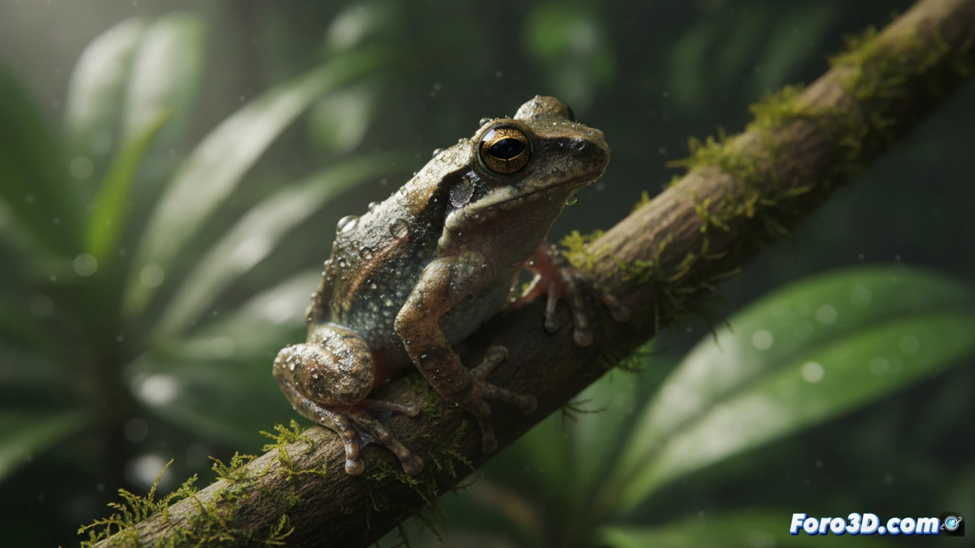
Technical workflow for microfauna 🐸
Modeling requires a high-precision approach. We start with photogrammetry of preserved specimens to capture the skin texture, granular and almost translucent. Then, in Blender, we retopologize the mesh to obtain a clean geometry of 25,000 polygons, ideal for real-time rendering. The key is in the scale: we include a reference asset, such as a 1 euro coin or a human fingertip, to contextualize the actual size of 8 mm. For animation, we develop a non-destructive rig that simulates jumping and abdominal breathing, synchronized with an ambient lighting cycle that mimics the humid understory of Madagascar.
The educational value of the infinitesimal 🔬
Beyond the technical challenge, this 3D model serves a crucial educational purpose. By allowing the user to rotate the frog and zoom in on its bone structure or its digital adhesive discs, access to science is democratized. In a virtual museum, the visitor can compare Mini mum with a common frog, visually understanding ecological specialization. It is not just a model; it is a tool to appreciate the hidden biodiversity that lives under leaves, demonstrating that the smallest detail deserves the highest graphical fidelity.
What 3D modeling techniques allow for accurately representing the anatomical structures of Mini mum, the world's smallest frog, for scientific visualization?
(PS: fluid physics for simulating the ocean is like the sea: unpredictable and you always run out of RAM)