The recent description of Phyllium gardabagusi in Indonesia (2023) has fascinated the scientific community due to its extreme mimicry. This leaf insect not only replicates the shape of a leaf but also integrates details such as herbivore bites, fungal spots, and irregular veins. For scientific visualization, it represents a technical challenge: capturing the transparency, organic texture, and natural deformation of its wings. Photogrammetry and high-resolution 3D scanning become essential tools for documenting this evolutionary deception.
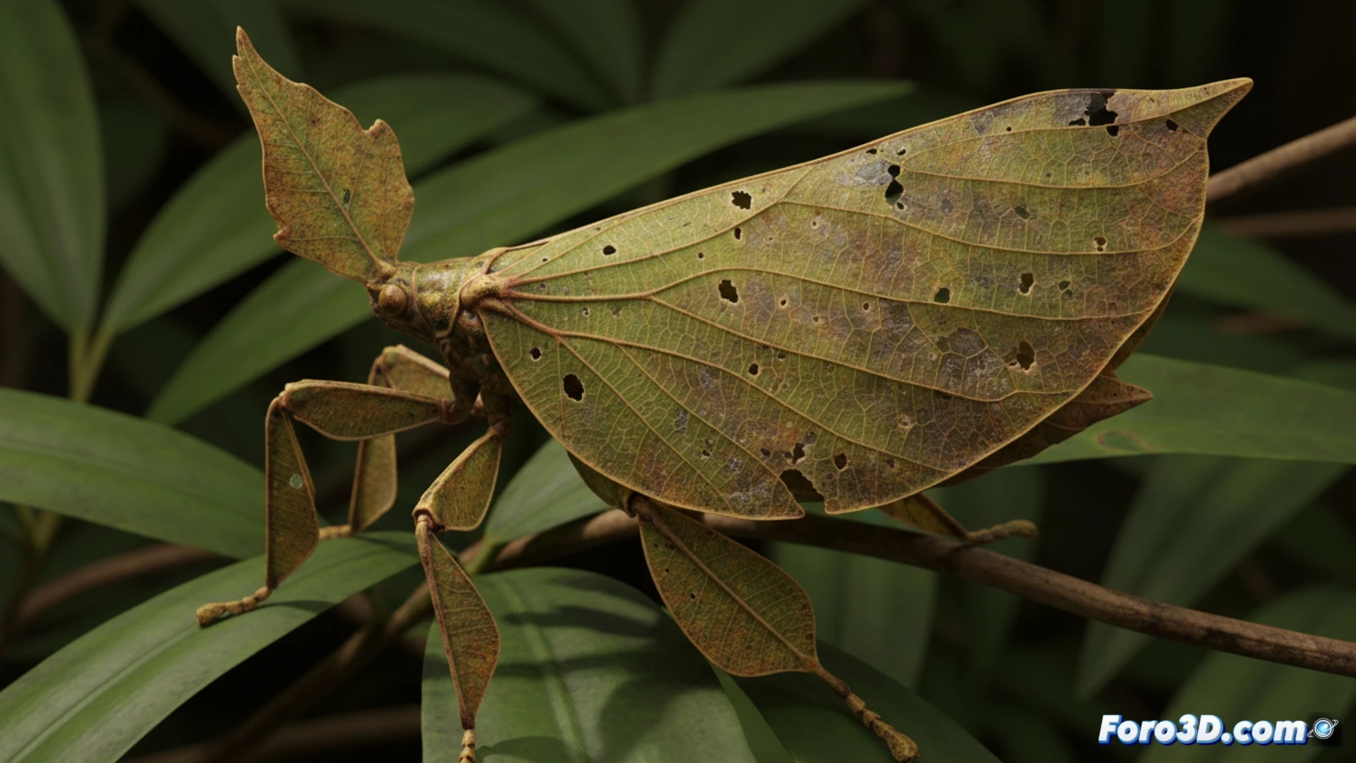
Photogrammetry and capture of biological textures 🧬
To accurately model Phyllium gardabagusi, a workflow combining multiple capture techniques is required. Cross-polarized photogrammetry allows recording the microtopography of its wings, including the depressions that mimic bites from real insects. The use of multispectral cameras helps isolate the chlorosis and necrosis patterns simulated by the insect. A critical step is capturing the body's translucency, achieved through polarized light scanning and subsequent subsurface scattering (SSS) simulation in rendering software. The resulting point cloud is processed in Blender or ZBrush to reconstruct the polygonal mesh, preserving the wrinkles and irregular edges that make the insect indistinguishable from a real leaf in a virtual museum.
Educational value and evolution in 3D 🌿
These three-dimensional models transcend simple illustration. By allowing rotation and zoom on the mimetic structures, researchers can analyze the coevolution between the insect and its plant environment. For virtual museums, a textured model of P. gardabagusi placed next to a leaf from its natural habitat (Cinnamomum) offers an interactive lesson in natural selection. The ability to decompose the model into layers (cuticle, tissue, venation) helps biology students understand how predatory pressure shapes form and color. Thus, scientific visualization not only documents the species but reconstructs the evolutionary process in an immersive digital environment.
What 3D scanning and parametric modeling techniques are most effective for capturing and reproducing the wing microstructures of Phyllium gardabagusi that enable its near-perfect mimicry of tropical leaf venation?
(PS: at Foro3D we know that even manta rays have better social bonds than our polygons)