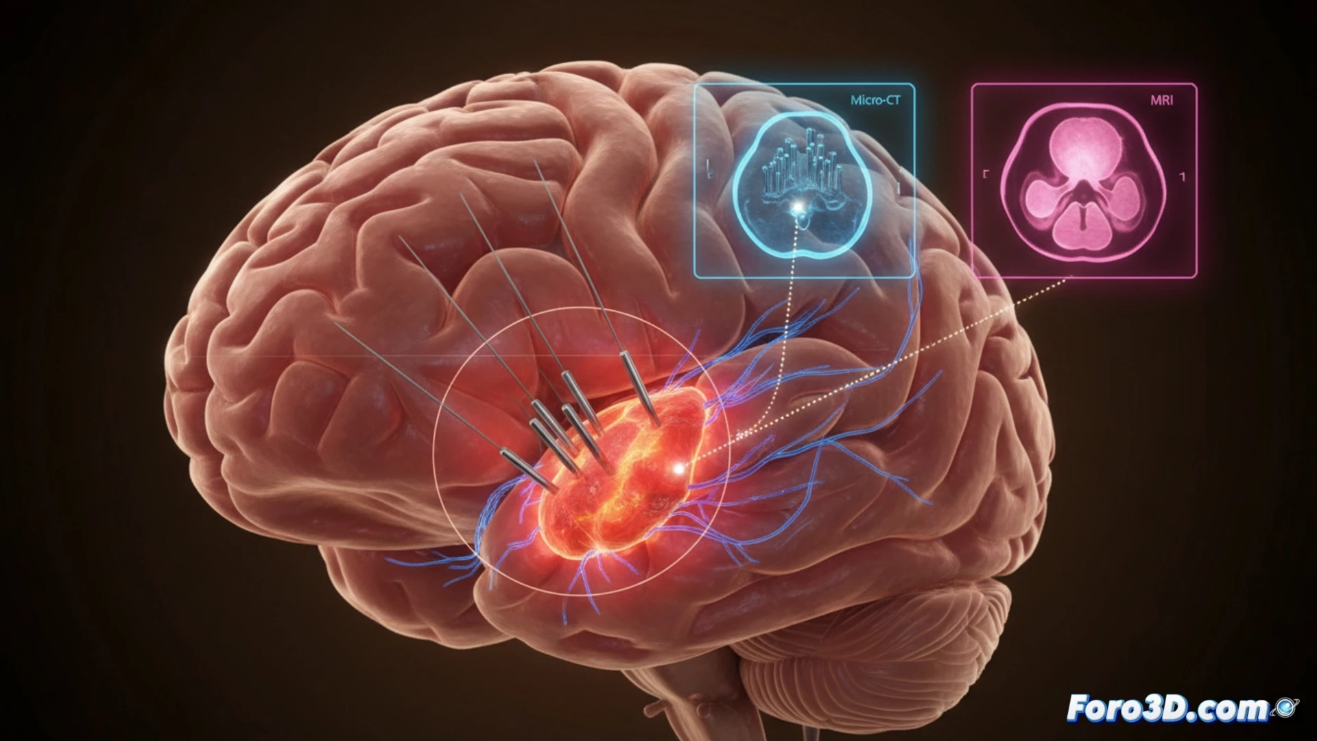
A patient with a next-generation neural implant experienced a sudden loss of motor control, despite the device's calibration software showing no anomalies. However, an advanced 3D pipeline, cross-referencing micro-CT and MRI data, revealed the real cause: the micro-electrodes had migrated 500 microns due to an undetected inflammatory reaction. This finding demonstrates the limitations of standard monitoring systems and the need for more precise diagnostic tools.
Workflow: Segmentation, fusion, and biomechanical simulation 🧠
The clinical team used Brainlab for surgical planning and initial fusion of high-resolution MRI images. Subsequently, in Materialise Mimics, a detailed segmentation of brain tissue and platinum-iridium electrodes was performed, allowing for an accurate 3D reconstruction. The micro-CT images provided the necessary resolution to visualize the exact position of each contact. Finally, the 3D model was exported to Ansys Biomechanic, where tissue behavior under a chronic inflammatory reaction was simulated. The simulation confirmed that the force generated by gliosis was sufficient to displace the electrodes, explaining the implant failure and the consequent loss of functionality.
Lessons for neural implant safety ⚠️
This case highlights an uncomfortable truth: current calibration algorithms are blind to subtle mechanical changes at the tissue-electrode interface. The integration of a 3D pipeline like the one described should become a post-operative standard for BCI implants. It is not just about detecting failures, but about predicting them through biomechanical simulations. Ignoring tissue dynamics is a risk that the next generation of neural implants cannot afford.
How can 3D detection of inflammation-induced BCI microelectrode migration improve precision in restoring motor signals after sudden loss of control in next-generation neural implants?
(PS: If you 3D print a heart, make sure it beats... or at least doesn't cause copyright issues.)