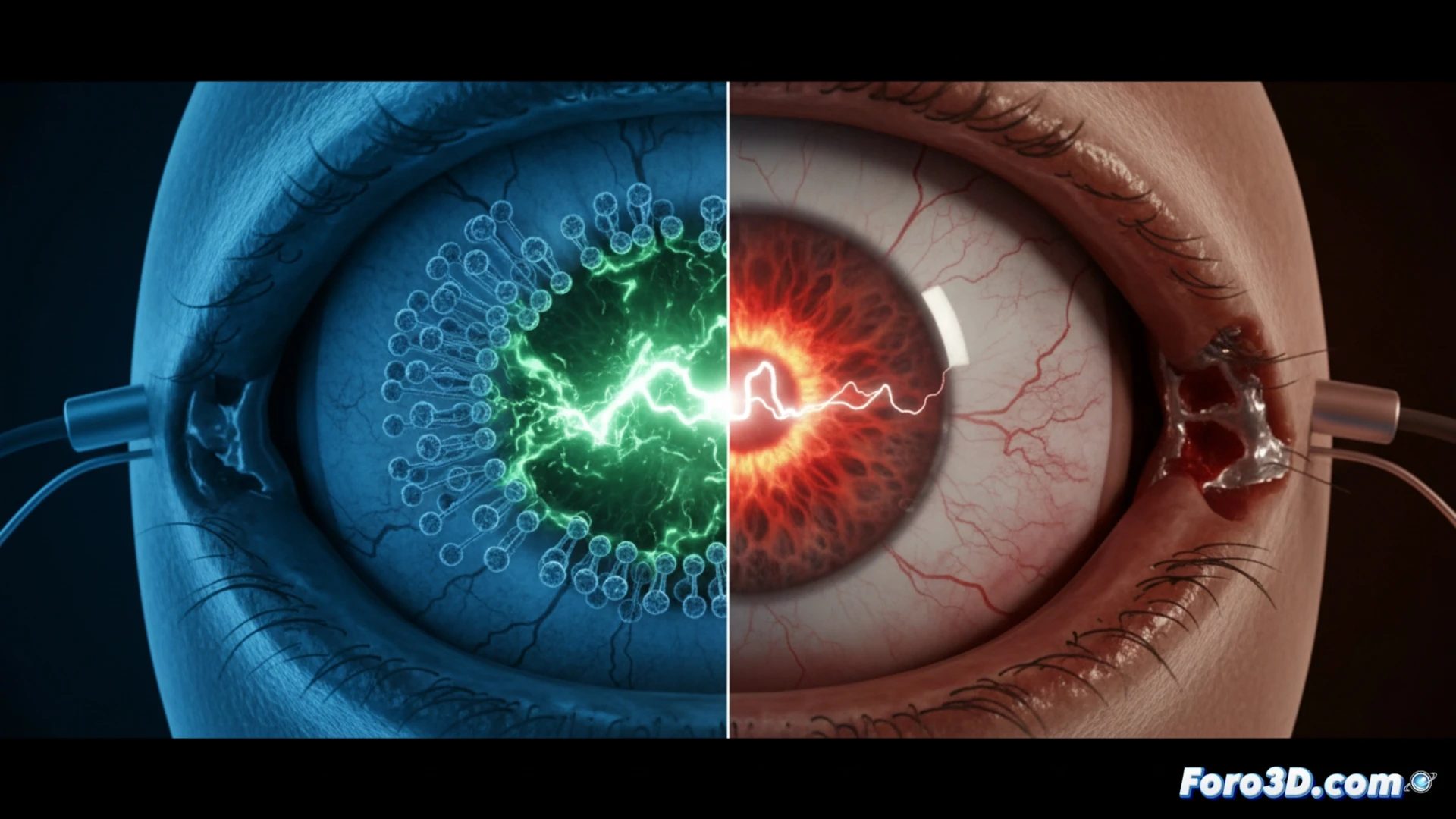
A patient suffered an internal burn after the activation of an artificial retina implant. Forensic analysis of the explanted device, using micro-CT and electromagnetic simulation, has allowed the failure mechanism to be reconstructed. The main hypothesis points to an entry of electrolyte-rich biological fluid, which caused an electric arc in the microelectrode array.
Forensic workflow: scanning, segmentation, and simulation 🔬
The process begins with a scan of the implant using high-resolution micro-CT, capturing the internal geometry of the sealed device. The DICOM data is imported into Materialise Mimics to segment cavities and potential fluid entry pathways. The resulting 3D model is transferred to Volume Graphics VGSTUDIO MAX, where structural integrity is analyzed and areas of degradation are identified. Finally, the mesh is exported to COMSOL Multiphysics, using the Bio-electromagnetism module to simulate electrolyte conductivity and predict the electric arc trajectory. The correlation between the simulated burn zones and those observed in the explant validates the short-circuit hypothesis.
Lessons for bioelectronic implant design ⚡
This case demonstrates that the combination of micro-CT and multiphysics simulation is essential for failure investigation in medical devices. The ability to visualize in 3D the interaction between biological fluids and microscopic circuits opens the door to more robust designs. Retinal implant manufacturers can now optimize seals and dielectric coatings, reducing the risk of electric arcs and improving long-term patient safety.
What micro-CT and electromagnetic simulation methodologies were used in the forensic analysis to identify the exact point of the short circuit in the retinal implant that caused the internal burn
(PS: and if the printed organ doesn't beat, you can always add a little motor... just kidding!)