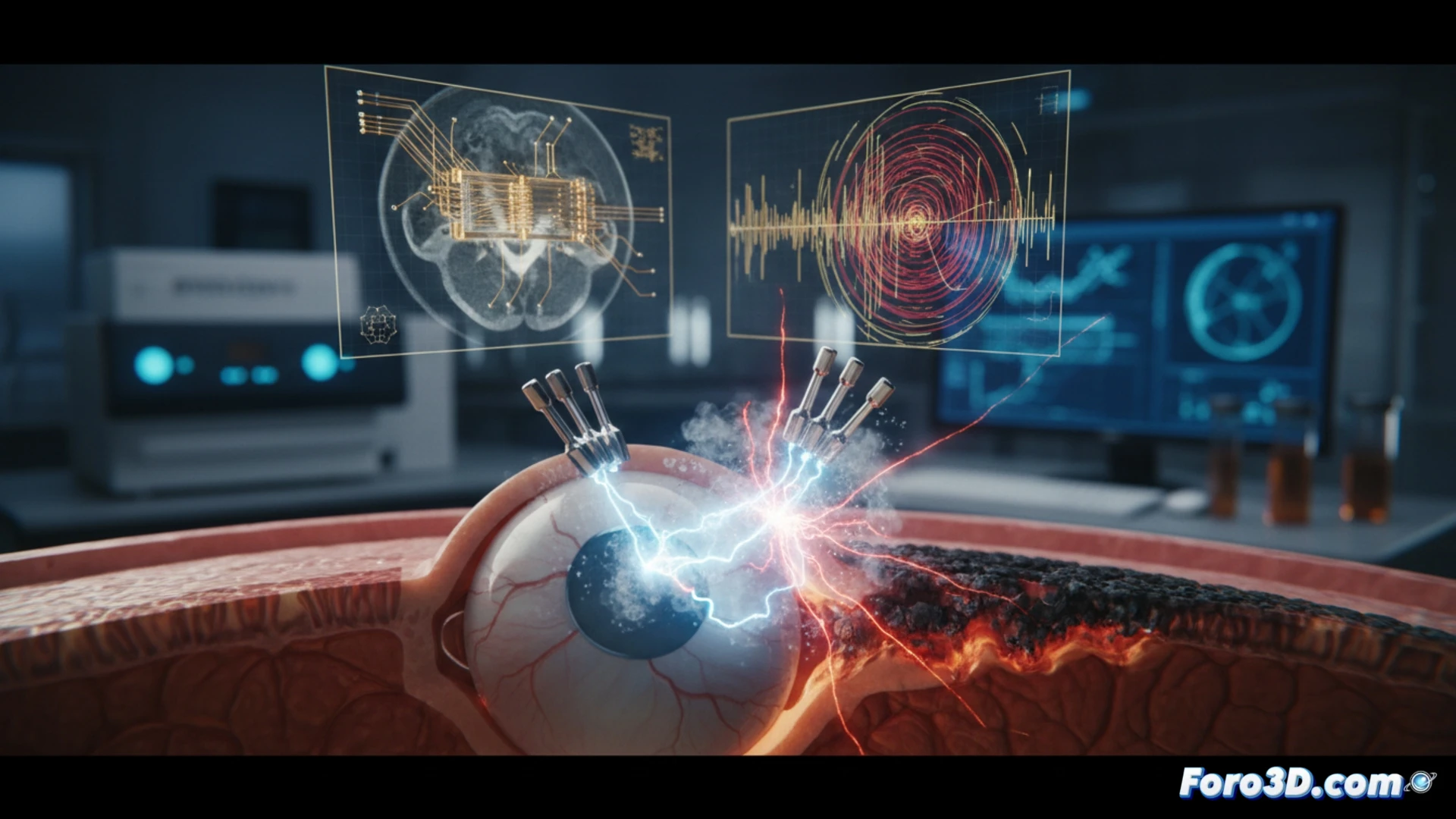
A patient suffered a severe internal burn when their artificial retina implant activated abnormally. The failure originated in the device's micrometric electrode array. To determine the cause, a forensic team applied micro-CT and electromagnetic simulation. The objective was to verify whether the ingress of biological electrolytes generated an electrical arc, damaging the surrounding tissue and compromising implant safety.
3D forensic workflow for implantable devices 🔬
The analysis began with scanning the explanted implant using micro-CT in Volume Graphics VGSTUDIO MAX software. The reconstructions revealed microcracks and carbonization zones in the insulating layer. Using Materialise Mimics, the volumes of infiltrated biological fluid were segmented. This data was exported to COMSOL Multiphysics to simulate the electric field in the Bio-electromagnetism module. The simulation confirmed that bodily electrolytes closed the circuit between adjacent electrodes, generating a high-temperature arc that burned the retina.
Lessons for designing safer implants ⚡
This case demonstrates that the combination of micro-CT and 3D simulation is crucial for investigating failures in medical devices. Identifying fluid entry points allows for redesigning hermetic seals and electrode layouts. The industry must adopt these virtual validation methods to predict short circuits under physiological conditions. Only then can future incidents be avoided and the reliability of electronic implants in the human body be guaranteed.
How could 3D finite element simulation applied to micro-CTs of retinal implants predict dielectric failure points before clinical validation, thereby avoiding risks of internal short circuits like the one reported?
(PS: and if the printed organ doesn't beat, you can always add a little motor... just kidding!)