Fracture of a keratoprosthesis implant, or artificial cornea, represents a critical challenge in regenerative ophthalmology. Thanks to the combination of 3D micro-CT and biomechanical simulations with Materialise Mimics and ANSYS, it is possible to analyze the polymer-tissue interface with micrometric precision. This approach reveals how hydrolysis degradation and mechanical fatigue induced by constant blinking compromise the structural integrity of the device.
Biomechanical simulation of the polymer-tissue interface 🔬
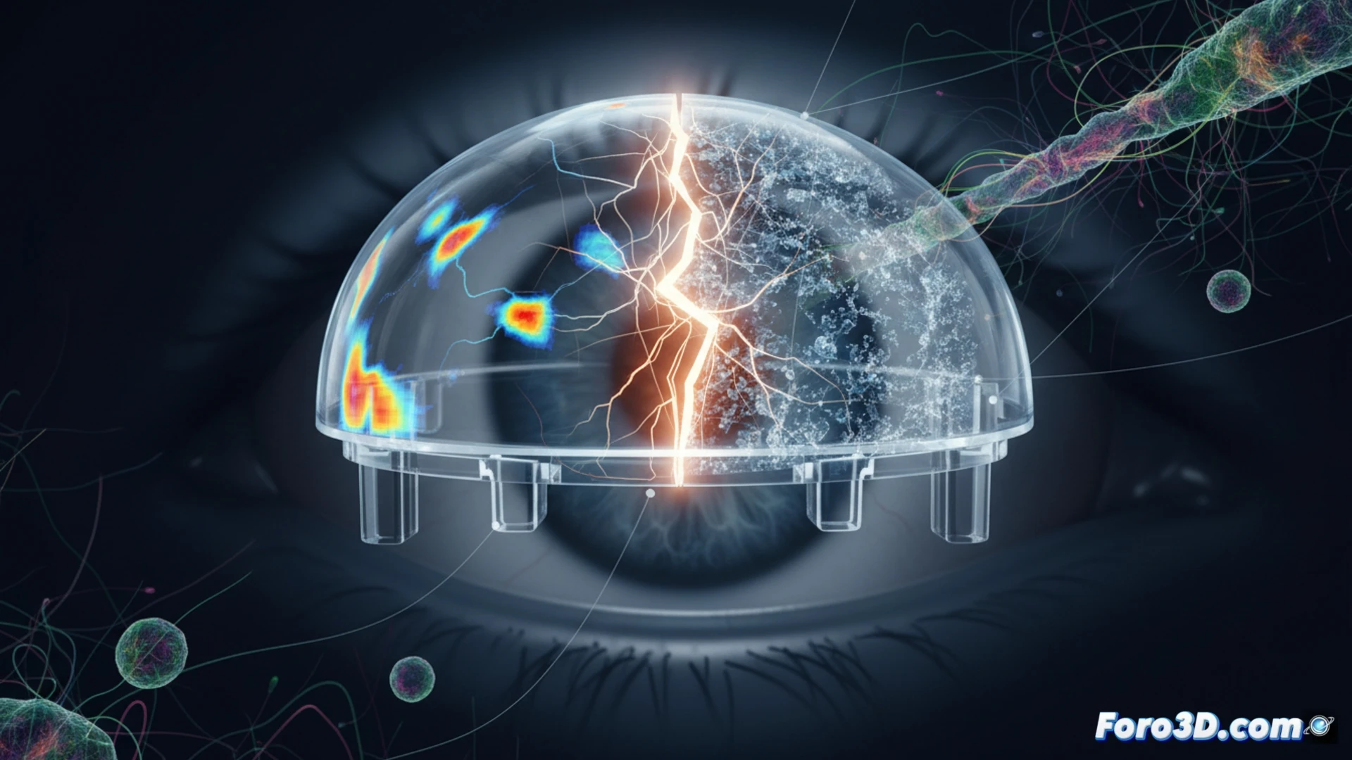
The technical workflow begins with image acquisition using confocal microscopy or micro-CT, processed in ZEISS ZEN 3D to segment the volume of the implant and surrounding corneal tissue. With Materialise Mimics, a three-dimensional model of the interface is reconstructed, identifying areas of detachment or microcracks. This model is exported to ANSYS Biomechanics, where cyclic loads simulating blinking pressure (approximately 15,000 blinks per day) are applied. The results show stress concentrations at the polymer edges, accelerating the hydrolysis of ester bonds in materials such as PMMA or hydrogel. Accumulated fatigue generates cracks that, without early detection, lead to complete implant fracture.
Towards more resistant ocular prostheses 💡
This analysis not only explains why current implants fail but also guides the design of new keratoprostheses. By correlating micro-CT data with fatigue simulations, engineers can modify the surface topography of the polymer to better distribute stresses or add bioactive coatings that resist hydrolysis. Integrating these 3D tools into the virtual prototyping phase will reduce failed clinical trials and improve the quality of life for patients with corneal blindness. Computational biomechanics is consolidating as a pillar in the validation of implantable medical devices.
It is possible that micro-CT revealed the exact location of the fracture in the keratoprosthesis, but how was that geometric information translated into a finite element model in ANSYS to predict failure propagation under physiological load?
(PS: and if the printed organ doesn't beat, you can always add a little motor... just kidding!)