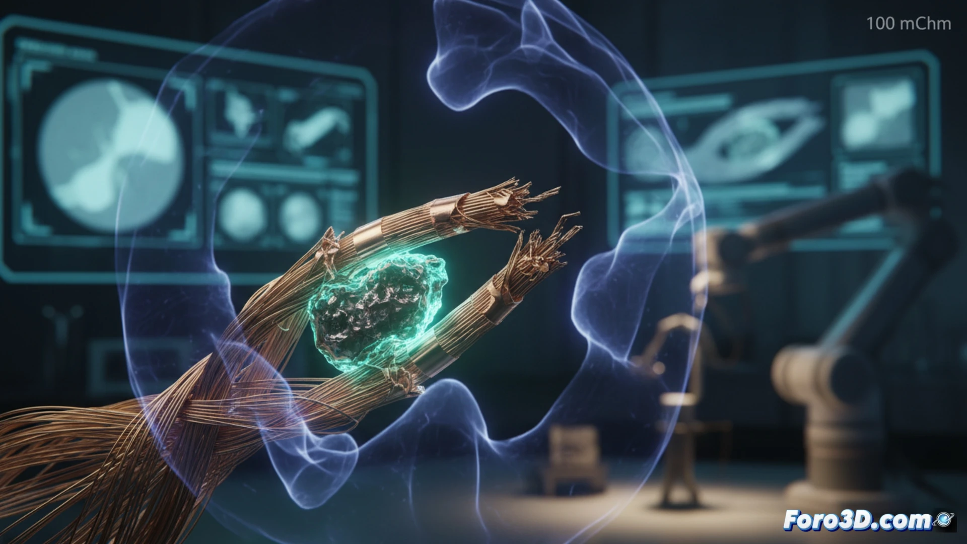
A Nitinol micro-gripper, designed for minimally invasive robotic surgery, fractured during an intervention, leaving a fragment inside the patient. The 100-micron wire, known for its shape memory, failed catastrophically. The forensic team turned to a non-destructive 3D analysis to determine whether the cause was a manufacturing defect or material fatigue.
3D Reconstruction and Fatigue Simulation in Nitinol 🛠️
Using VGSTUDIO MAX, the fractured wire was scanned with micro-CT, achieving submicron resolution that revealed a 5-micron titanium oxide inclusion. This particle, embedded during the drawing process, acted as a stress concentrator. The 3D model was imported into Ansys, where a shape memory deformation cycle was applied. The finite element simulation demonstrated that the inclusion generated a local stress 40% higher than the material's fatigue limit, initiating the crack that led to the fracture.
Lessons for Medical Device Manufacturing 🔬
This case underscores that, even with advanced materials like Nitinol, the purity of the manufacturing process is critical for patient safety. The combination of micro-CT and Ansys simulation not only identified the root cause but also allowed for proposing stricter quality control in alloy ingots. Visualization in Blender facilitated communication of the failure to the clinical team, demonstrating the value of 3D analysis in biomedical forensic engineering.
How can micro-computed tomography improve the design of Nitinol micro-grippers to prevent embrittlement induced by cyclic stress in robotic surgery
(PS: If you 3D print a heart, make sure it beats... or at least doesn't cause copyright issues.)