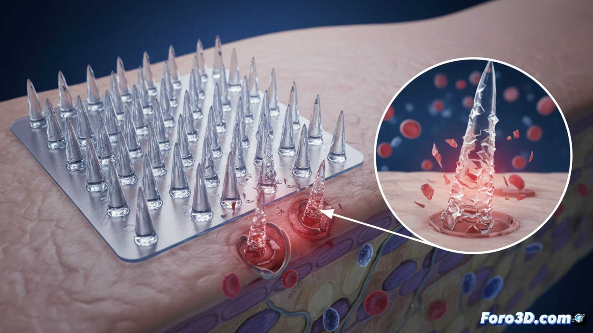
A batch of needle-free vaccination patches has caused adverse reactions in patients. Initial suspicion pointed to the geometry of the micro-needles, barely 200 microns long. The quality control team turned to a 3D workflow to discover the root cause: microscopic burrs that, instead of penetrating the skin cleanly, tore the tissue.
Technical workflow: from profilometry to simulation 🔬
The process began with high-resolution scanning using a Keyence VK Analyzer confocal microscope, generating topographic profiles of the tips. With this data, the point clouds were imported into VGSTUDIO MAX for micro-geometry analysis. The software allowed comparing each needle against the original SolidWorks CAD design, revealing critical shape deviations. Penetration simulation in a virtual tissue model confirmed that the burrs, less than 10 microns in size, generated shear forces exceeding the dermis's breaking limit, causing micro-tears.
Lessons for medical device manufacturing 🏥
This case underscores the need to integrate 3D quality control into the production of biomedical devices. An injection molding error, imperceptible to the naked eye, compromised the safety of an entire batch. The combination of profilometry, volumetric analysis, and simulation not only identified the failure but also establishes a protocol to validate the integrity of any micro-needle patch before distribution.
Since the initial suspicion pointed to the defective geometry of the micro-needles detected through 3D analysis, which microscopic design parameter was primarily responsible for the micro-needles failing to penetrate the epidermal layer correctly without causing damage?
(PS: and if the printed organ doesn't beat, you can always add a little motor... just kidding!)