The discovery of the blood comb jelly (Lampocteis sp.) on Chilean seamounts during 2024 has opened a fascinating window for scientific visualization. This ctenophore, of an intense ruby color, possesses rows of cilia that emit a unique bioluminescence. For 3D graphics specialists, it represents a technical challenge and an opportunity to recreate the interaction between light and semi-transparent tissue in a deep-sea environment.
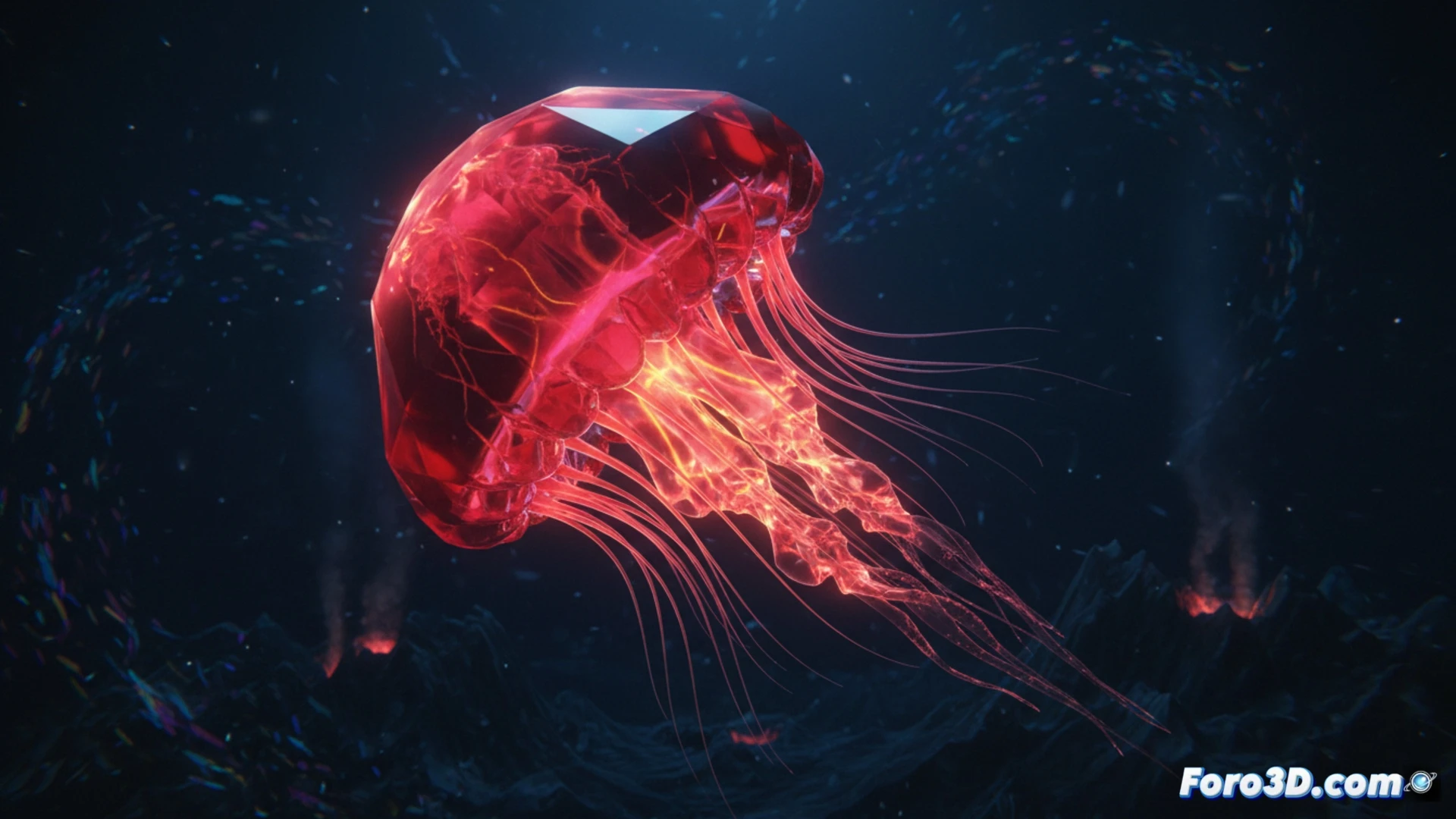
Photorealistic Modeling and Simulation of Bioluminescent Cilia 🌊
Developing a 3D model of this species requires a focus on subsurface scattering (SSS) to capture the deep ruby tone of its gelatinous body. The geometry must be organic, with a mesh that allows smooth deformations to simulate propulsion. The critical point is the animation of the eight rows of cilia; here, a particle system with point emitters can recreate the synchronized light flashes. It is recommended to use a rendering engine that supports volumes for water and oceanic fog, and procedural textures to avoid repetitions in translucency. HDRI lighting from a bathyal environment helps integrate the model into an interactive scientific documentary.
The Value of Biological Precision in Science Communication 🔬
Beyond technical realism, this 3D model serves a crucial educational function. It allows biologists and virtual museums to display the anatomy of a living being that is nearly impossible to observe in captivity. By comparing its cilia structure with other comb jellies, key evolutionary differences can be visualized. The challenge for the 3D artist is not to sacrifice scientific accuracy for visual impact, achieving a balance that does justice to the beauty of this creature from the Chilean seamounts.
What software do you recommend for visualizing animal tracking data?