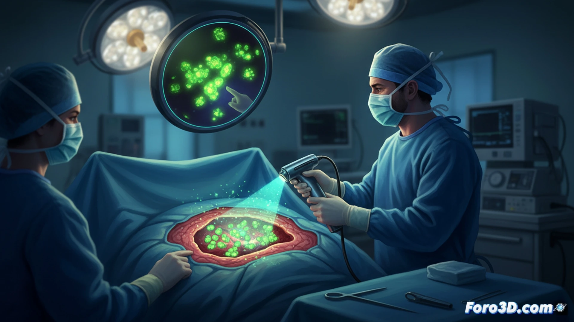
Oncological surgery faces a critical challenge: distinguishing healthy tissue from cancerous tissue in real time. Lumicell Swift, an FDA-approved system, addresses this problem through fluorescence molecular imaging. After injecting an agent that lights up upon contact with tumor cells, the surgeon scans the surgical bed with a handheld probe, detecting residual foci invisible to the human eye.
Technical architecture of the intraoperative fluorescence system 🔬
The system consists of a fluorescent agent (pegulicianine) and a portable imaging device. Pegulicianine is activated by an enzyme present in the tumor microenvironment, emitting light in the near-infrared spectrum. The detection probe integrates a high-sensitivity CMOS sensor and optical filters that isolate the fluorescent signal from healthy tissue. A real-time analysis software processes the data and generates a heat map overlaid on the surgical field image, guiding the surgeon to suspicious areas with submillimetric precision.
Implications for 3D surgery and oncological precision 🎯
Lumicell Swift aligns with trends such as 3D surgical navigation and printed anatomical models. While these tools offer preoperative planning, intraoperative fluorescence closes the loop by providing real-time verification. The result is a drastic reduction in reoperation rates, as it allows complete tumor removal in a single operation, transforming the standard of care in breast, prostate, and other solid tumor surgeries.
How does the surgical community evaluate the impact of Lumicell Swift's intraoperative fluorescence on reducing positive margins during complex oncological procedures?
(PS: and if the printed organ doesn't beat, you can always add a little motor... just kidding!)