Scientists have captured in three dimensions how T lymphocytes destroy cancer cells. Using advanced microscopy, they observed in real time the formation of an immunological synapse, the point of contact where the immune system recognizes and attacks the tumor. This breakthrough allows seeing details never before recorded of this cellular battle.
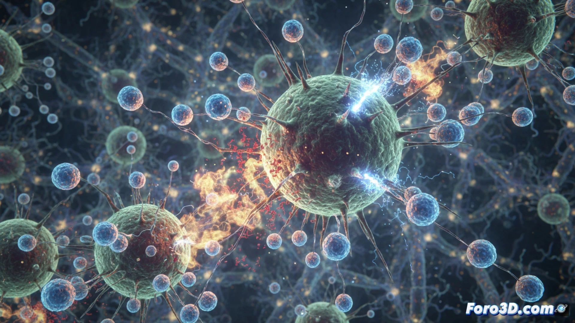
Next-generation microscopy reveals the immunological synapse 🔬
The team used fluorescence and super-resolution microscopy techniques to film the interaction between the T lymphocyte and the tumor cell. The images show how the lymphocyte deploys receptors and releases cytotoxic molecules in an organized space called the immunological synapse. This process, previously only inferred, is now seen in three-dimensional detail, helping to understand failures in current therapies and to design new treatments.
Killer cells in slow motion: the ultimate selfie 📸
Seeing a T lymphocyte corner a cancer cell in 3D is fascinating, but also a bit unsettling. It's as if the immune system has learned to take a selfie while working. Of course, the tumor cell doesn't pose for the photo; it dies without glamour. Science advances, and soon we'll see tumors on TikTok, though no one will give them a like.