A scientific team has managed to observe in three dimensions how a T lymphocyte destroys a cancer cell. The study, published in Science, shows the process in real time. T lymphocytes, known as killer cells, identify the tumor, adhere to it, and release toxic granules. This is the first time the immune system's mechanism of action against cancer has been seen with such clarity.
The immunological synapse in high resolution 🧬
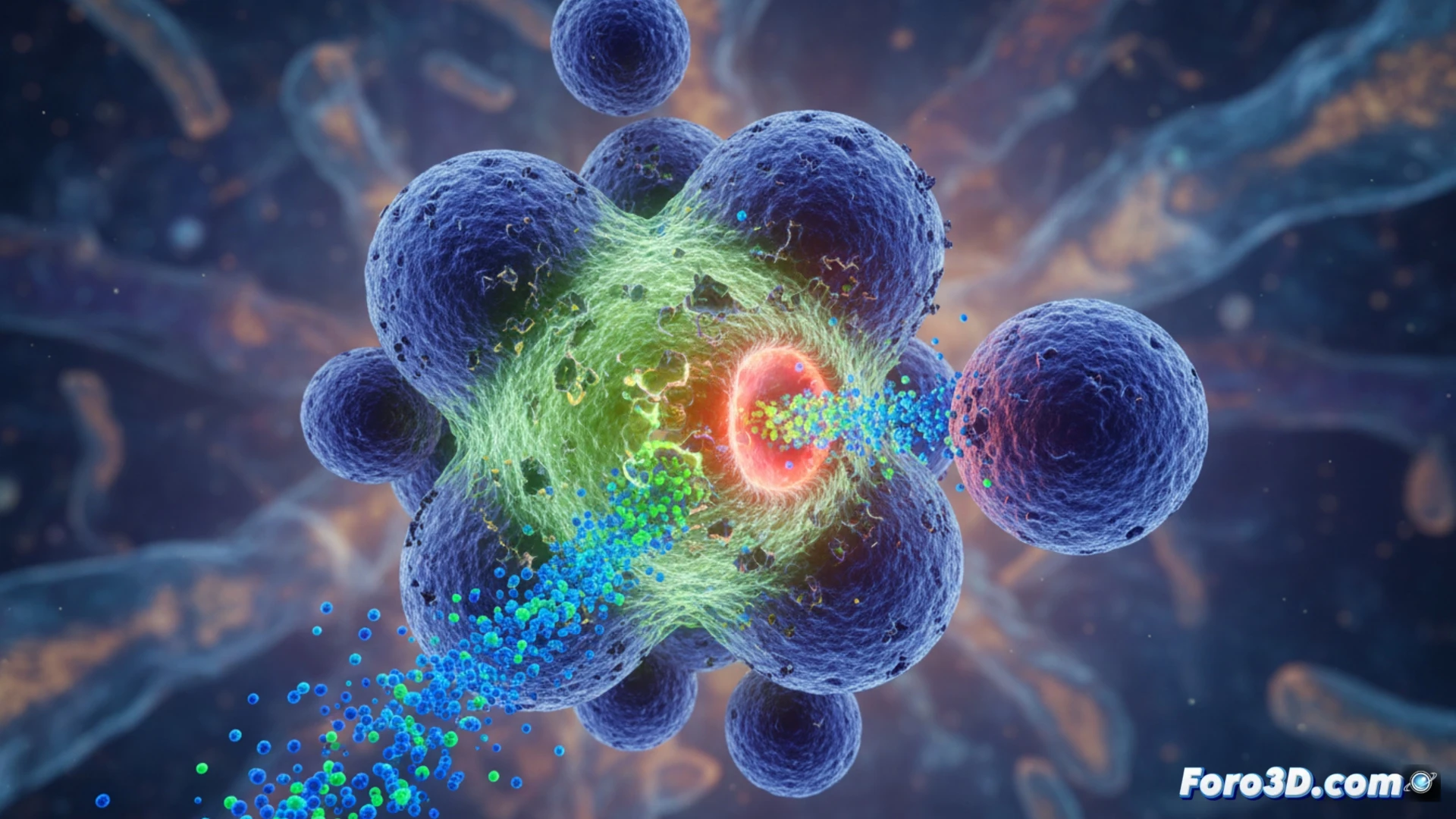
The new 3D imaging technique allows each step of the attack to be followed. First, the T lymphocyte recognizes the tumor cell and forms an immunological synapse, a specialized contact zone. Then, it releases lethal enzymes at a precise point. Scientists managed to capture the formation of pores in the cancer cell membrane and the entry of toxins. This level of detail opens the door to understanding why some tumors resist the attack and how to improve immunotherapies.
The T lymphocyte: a killer with GPS and laser precision 🎯
Watching a T lymphocyte in action is like witnessing a highly precise surgical execution. There are no stray bullets or collateral damage: the killer selects its target, locks on, and fires its lethal payload right where it hurts. Meanwhile, the surrounding healthy cells watch the show unfazed. If T lymphocytes had a talent agency, they would already be flooded with Hollywood offers for hitman roles.