Professional tennis is not just an elite sport; it is a high-risk biomechanical profession. Repetitive movements, explosive changes of direction, and varying surfaces make tennis players a case study for occupational injury prevention. From tennis elbow to hamstring tears, each stroke accumulates stress on the musculoskeletal system. This is where 3D technology ceases to be a luxury and becomes an essential diagnostic and corrective tool.
Motion capture and biomechanical modeling 🎾
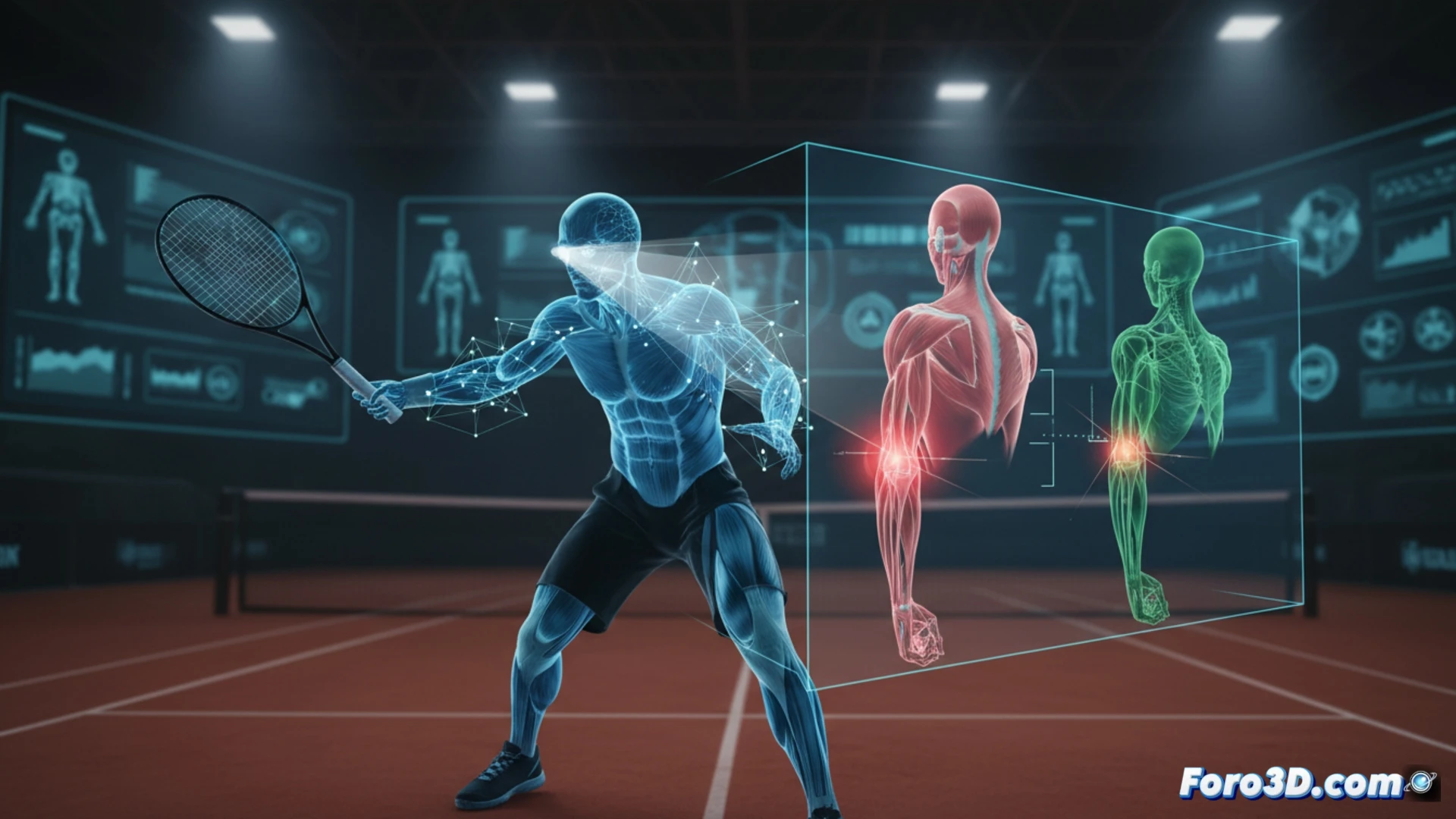
Motion capture (mocap) with inertial sensors or infrared cameras allows real-time digitization of the tennis player's kinematics. By modeling the body in a 3D environment, physiotherapists can isolate variables such as wrist angle on the backhand or shoulder rotation on the serve. For example, a simulation may reveal that a player generates elbow hyperextension due to poor trunk alignment, increasing the risk of epicondylitis. By overlaying the athlete's technique with an optimal model, millimeter deviations are identified that, repeated thousands of times, lead to knee tendinitis or ankle sprains from unstable lateral supports.
Preventing wear, not just treating injury 🛡️
The true revolution lies in moving from reactive rehabilitation to predictive prevention. With 3D simulations, a coach can adjust footwork on a slippery court before a fall occurs, or modify the kinetic chain of a stroke to reduce lumbar fatigue. This approach, combining biomechanical data with virtual reality, allows professional tennis players to extend their careers without accumulating the typical occupational wear and tear. 3D technology does not just analyze injuries: it prevents them.
How can 3D motion capture identify muscle fatigue patterns in professional tennis players before they become chronic injuries?
(PS: at Foro3D we know that a simulated penalty in 3D always goes in... unlike in real life)