3D technology is transforming prehospital care. Paramedics can now use printed anatomical models to practice complex procedures before facing a real patient. A clear example is the simulation of difficult airways: printing an exact replica of a patient's trachea allows for risk-free emergency intubation practice. Necessary programs include medical segmentation software like 3D Slicer and modeling with Blender or Meshmixer.
From scanner to ambulance: the technical workflow 🛠️
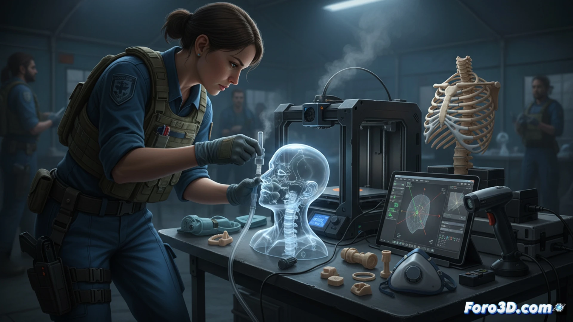
The process begins with a CT scan or MRI of the patient, whose DICOM files are processed in 3D Slicer to isolate the target anatomy. Then, the model is refined in Blender to remove artifacts and simplify geometries. Finally, it is exported to STL format and printed on an FDM or SLA printer with biocompatible materials. Programs like PrusaSlicer or Cura configure the printing parameters. The result: a tactile simulator that the paramedic can use to train critical maneuvers such as cricothyrotomies or intraosseous accesses.
When you print a lung and the patient breathes by miracle 😅
The best part is that you can have a printed lung in your backpack, but the real patient will still be coughing as if you had given them a mint gum. Paramedics now carry 3D pieces instead of carrying more gauze. The only problem is that if you print the trachea wrong, the patient stops breathing and you're left with an anatomical paperweight. That said, at least you can explain to the boss that the error was the software's fault, not yours.