3D technology offers practical tools for the midwifery profession, facilitating the visualization of fetal and pelvic anatomy. With printed models or virtual simulations, complications during childbirth, such as shoulder dystocia or abnormal positions, can be anticipated. This improves communication with the patient and the planning of the medical team.
Anatomical models and segmentation software 🧬
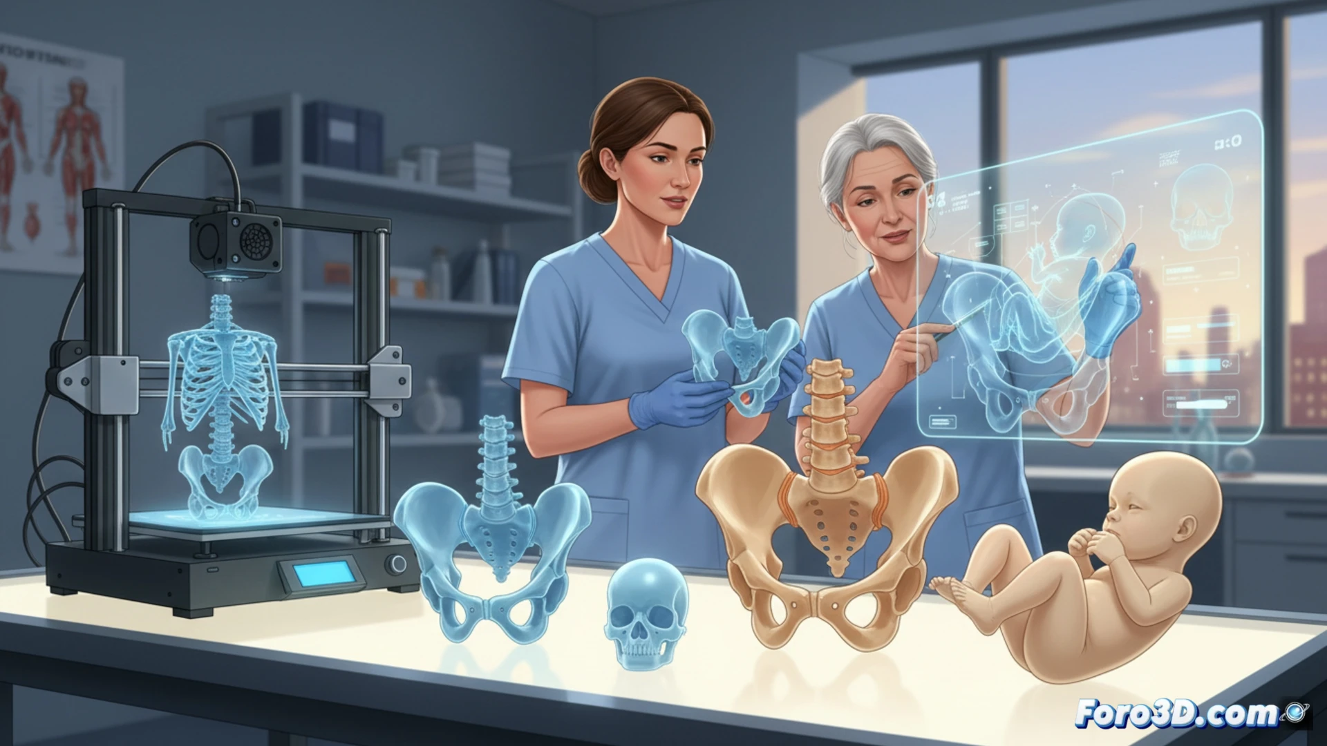
To create a 3D model of the fetal and maternal pelvis, an MRI or 3D ultrasound is used as a starting point. The 3D Slicer software allows for segmenting bones and tissues, exporting STL files. Then, Blender is used to clean the mesh and add markers. Printing is done with PLA filament on a printer like the Creality Ender 3. The result is a tactile model for practicing maneuvers such as Leopold's.
Childbirth with a printer: instruction manual included 🤖
Now midwives can print the baby before it arrives. If it doesn't come out like in the ultrasound picture, they can always blame the bed calibration. Of course, don't try using PLA for the umbilical cord; nature already designed that and doesn't need supports. At least the printer doesn't ask for an epidural.