3D technology is transforming veterinary medicine by enabling the planning of complex surgeries with exact patient models. A dog with a complicated pelvic fracture can be operated on using a printed surgical guide, reducing time and errors. Programs like 3D Slicer for segmenting scans and Fusion 360 for designing implants are key tools in this change.
Digital modeling and biomodels for veterinary surgery 🐾
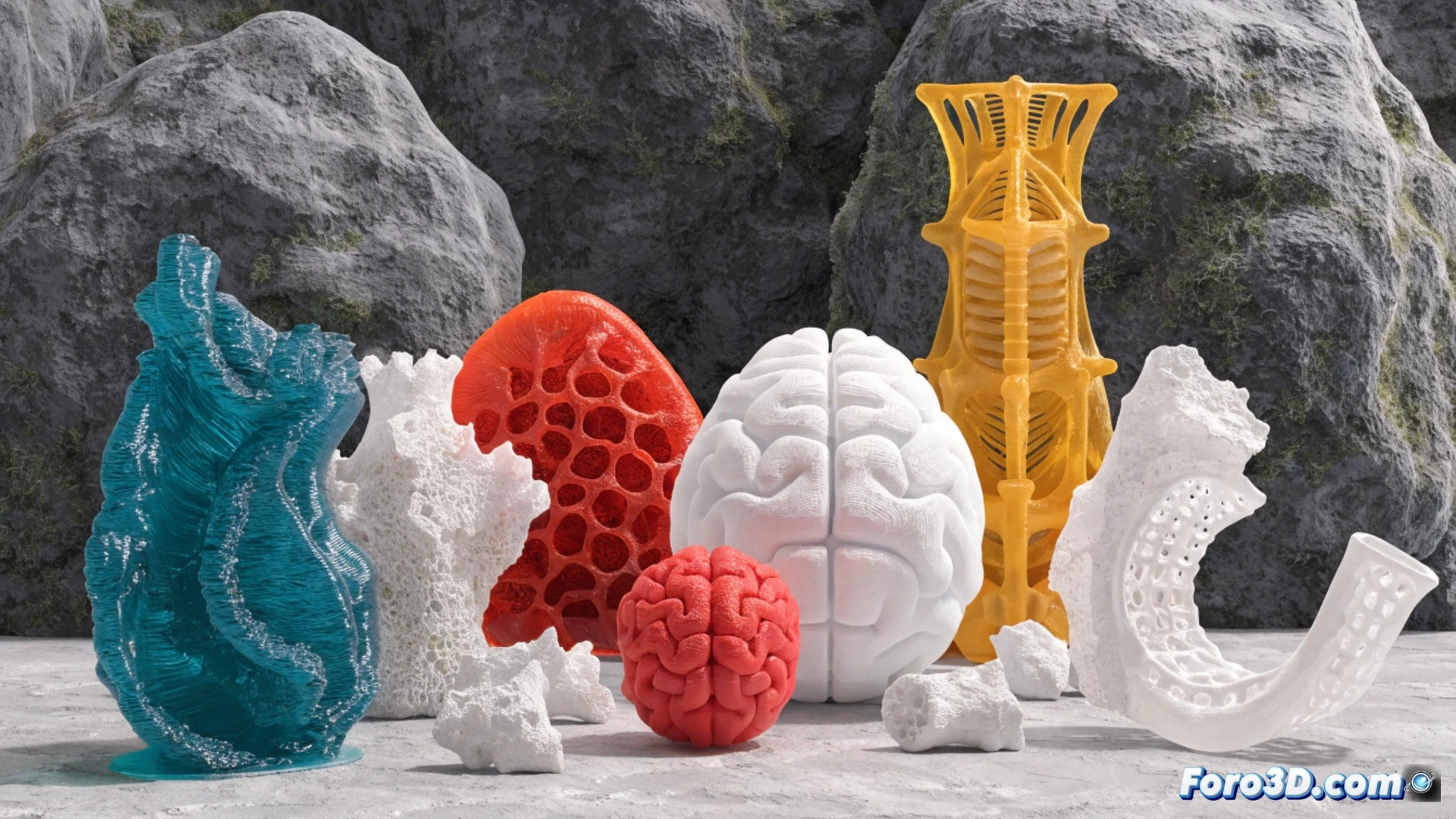
The process begins with a CT scan or MRI of the animal. The DICOM file is processed in 3D Slicer or InVesalius to isolate the damaged bone or tissue. Then, in Meshmixer, the mesh is cleaned and the prosthesis or splint is designed. Printing in PLA or titanium allows for the creation of custom implants. This avoids stocking standard sizes and makes it easier to adapt to breeds with atypical anatomies, such as greyhounds or bulldogs.
When the cat refuses to cooperate with the scanner 😼
The biggest problem is not the software, but convincing a cat to stay still during the CT scan. I have seen colleagues use fleece socks and tuna as a sedative. And if the 3D model of the femur comes out crooked because the patient moved, the scan has to be repeated. In the end, technology advances, but feline resistance remains the toughest technical challenge.