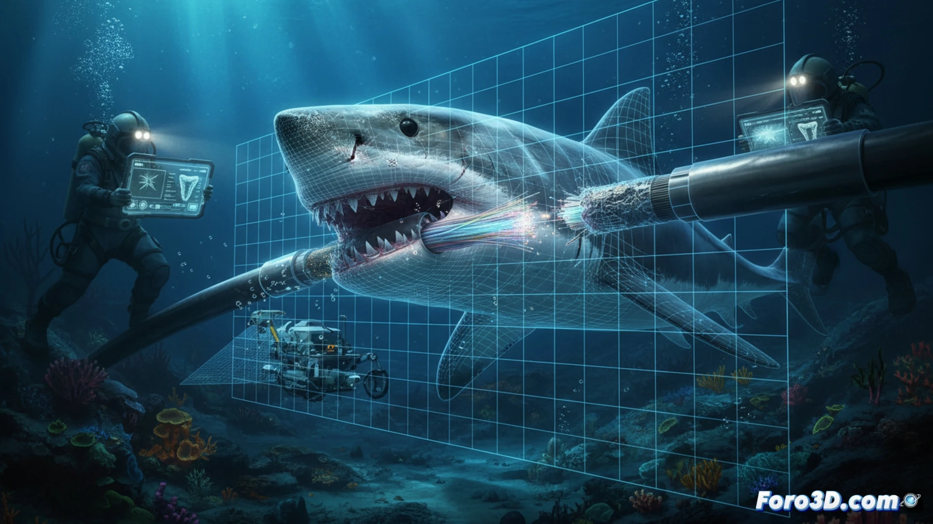
A transatlantic fiber optic cable goes out of service with no apparent cause. Engineers deploy an ROV equipped with high-resolution cameras to inspect the damage. Bite marks on the polyethylene sheath are evident, but the key question remains open: which marine species caused the break and whether the internal fibers survived the attack. The answer is not in the ocean, but in a three-dimensional model generated by underwater photogrammetry.
Forensic reconstruction with Agisoft Metashape and MeshLab 🦈
The process begins with capturing hundreds of images of the damaged segment, taken from different angles by the underwater vehicle. These images are processed in Agisoft Metashape to generate a dense point cloud and a high-fidelity polygonal mesh of the bitten area. The model is exported to MeshLab, where smoothing filters are applied and false color depth maps are calculated. These maps reveal the exact penetration of the teeth into the polyethylene, allowing measurement of whether the damage reached the Kevlar layer or the optical fibers themselves. The three-dimensional dental imprint is compared with databases of shark jaws and other marine predators. The morphology of the marks, especially the spacing between incisors and the curvature of the dental arch, points directly to a specific species, such as the blue shark or the mako, ruling out attacks by cetaceans or swordfish.
Implications for engineering and marine biology 🔬
This workflow demonstrates that 3D scientific visualization is not only useful for documentation but also for making critical decisions. Engineers confirm that, although the outer sheath is perforated, the internal optical fibers remain intact, avoiding costly cable replacement. For marine biologists, the model allows studying predator behavior without the need to capture them. The logical next step is to simulate the attack angle and bite force in Blender using soft body dynamics, closing the loop between remote observation and underwater biomechanics.
How point clouds generated by 3D photogrammetry are processed and analyzed to differentiate shark bite marks from other mechanical damage on submarine cables
(PS: modeling manta rays is easy; the hard part is making them not look like floating plastic bags)