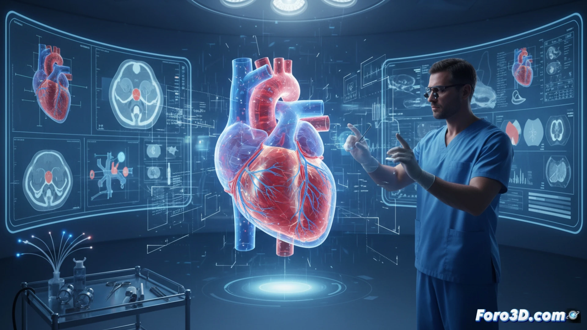
3D technology is transforming cardiology by allowing visualization of cardiac anatomy with millimeter precision. Three-dimensional models, created from CT scans or MRIs, help plan complex surgeries and simulate interventions before touching the patient. A clear example is the reconstruction of heart valves to practice their repair.
Key programs for modeling the heart 🛠️
To work with these models, programs like Mimics or 3D Slicer are used, which process DICOM images and generate STL files. Then, software like Blender or Meshmixer allows editing geometries to simulate cuts or implants. 3D printing with flexible materials (such as TPU) reproduces soft tissues, giving the cardiologist a physical prototype to rehearse complex procedures before the actual surgery.
When the heart beats in the printer 🖨️
Now cardiologists can hold a heart in their hand without having to ask anyone for it. Literally. They print it, look at it, rotate it, and even operate on it for practice before the big day. Of course, if the printer fails in the middle of the night, the patient will have to wait until maintenance is complete. Technology advances, but the classic filament jam remains the number one enemy of cardiac health.