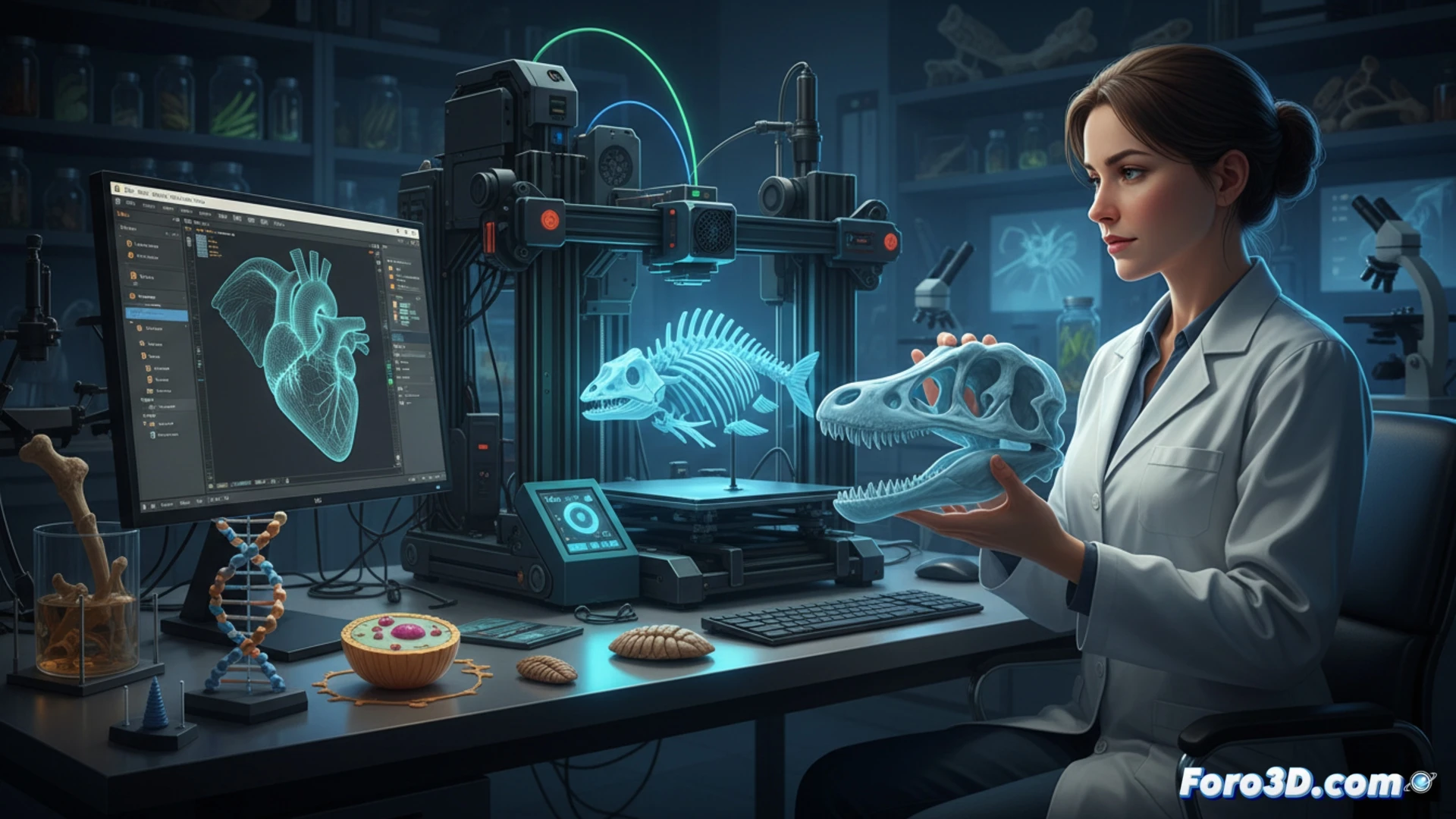
3D technology allows biologists to recreate anatomical models, ecosystems, and fossils with millimeter precision. A clear example is the replication of skulls of extinct species to study their biomechanics without damaging the original. Programs like Blender, Meshmixer, and 3D Slicer are basic tools for processing CT scans and generating pieces ready for printing.
Programs and workflow for the digital biologist 🧬
To go from a CT scanner to a physical piece, 3D Slicer is used to segment bones or tissues. Then, Blender cleans the mesh and corrects imperfections. Finally, Meshmixer or PrusaSlicer prepare the file for the printer. These programs are free and allow adjusting scales, adding supports, or even simulating joints. The result is a tactile model that facilitates teaching and research.
The biologist and their collection of plastic bugs 🦗
Now any biologist can have an exact replica of a trilobite fossil on their desk, right next to their cold coffee. The best part is that they no longer have to borrow the original from the museum and sign a document worth more than their salary. If the print fails, they only lose filament, not a unique 500-million-year-old piece. Of course, they shouldn't think of using it to crack nuts.