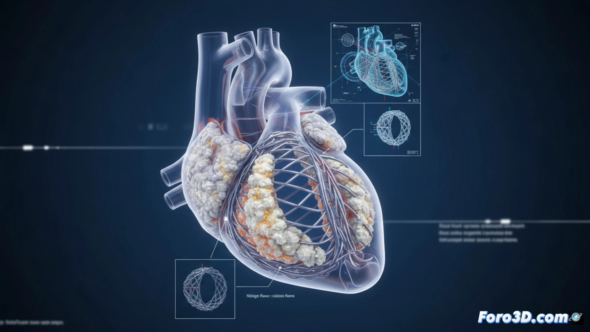
A patient suffers sudden stenosis in a catheter-implanted heart valve. To understand the cause, a 3D analysis is performed comparing the post-operative scan with the original Nitinol stent model. The objective is to determine whether the support geometry favored asymmetric calcium buildup. This clinical case demonstrates how reverse engineering tools can prevent implant failures.
Technical Workflow: From Mimics to Ansys 🛠️
The process begins with Materialise Mimics, where DICOM images from the post-implant CT scan are segmented to isolate the stent and calcified areas. Then, in GOM Inspect, this real geometry is superimposed against the original CAD design of the device. Chromatic deviation reveals local deformations of the Nitinol. Finally, Ansys Biomechanic simulates residual stresses in the stent, correlating high-deformation zones with calcium deposits. This workflow allows identifying whether implant asymmetry is a critical biomechanical factor.
Lessons for Prosthetic Valve Design 💡
The analysis shows that the differential stiffness of Nitinol can generate turbulent microflows that precipitate calcification. The 3D comparison between the ideal design and the post-operative reality is vital for the industry. It is not enough for the stent to be flexible; its expansion pattern must ensure homogeneous load distribution. Biomechanical simulation thus becomes a quality filter for future generations of TAVI valves, reducing the risk of restenosis.
How does the asymmetric distribution of calcium influence the deformation of the nitinol stent during post-TAVI 3D analysis, and what implications does it have for predicting sudden valvular stenosis?
(PS: If you 3D print a heart, make sure it beats... or at least doesn't cause copyright issues.)