Withings has introduced BeamO, a pocket-sized device that unifies a thermometer, electrocardiograph, oximeter, and digital stethoscope. This advancement allows users to perform basic cardiac and pulmonary check-ups without leaving home. However, the true potential of this tool is unlocked when we integrate its data with 3D visualization technologies. We analyze how volumetric modeling can transform these readings into interactive anatomical maps, improving telemedicine and early diagnosis.
3D Visualization of Integrated Physiological Signals 🫀
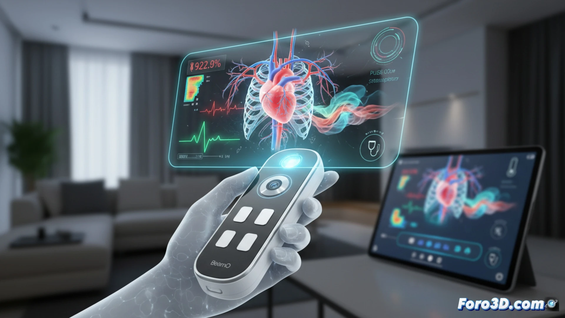
The data flow from BeamO can be represented in real-time using a three-dimensional model of the thorax. Electrocardiograph (ECG) signals are projected as depolarization vectors onto a 3D heart, while the oximeter and digital stethoscope generate point clouds that simulate lung expansion and respiratory sounds. This overlay allows physicians to identify anomalous patterns, such as murmurs or arrhythmias, by correlating acoustics with organ geometry. Additionally, interactive infographics can show the path of blood flow based on heart rate and oxygen saturation, offering a volumetric view that a 2D monitor cannot match.
The New Standard for Preventive Telemedicine 🏥
The combination of compact hardware like BeamO with 3D digital twins of organs marks a before and after in remote care. By modeling stethoscope data in a three-dimensional lung, the specialist can evaluate sound distribution in real-time, detecting areas of consolidation or pleural effusions without the need for an ultrasound. This symbiosis between portable sensors and anatomical rendering not only empowers the patient but also redefines the limits of preventive medicine from home.
Considering that BeamO integrates traditional sensors into a portable format, how could the incorporation of a low-cost 3D scanner in future versions of this device transform the early detection of structural cardiac anomalies at home?
(PS: and if the printed organ doesn't beat, you can always add a little motor... just kidding!)