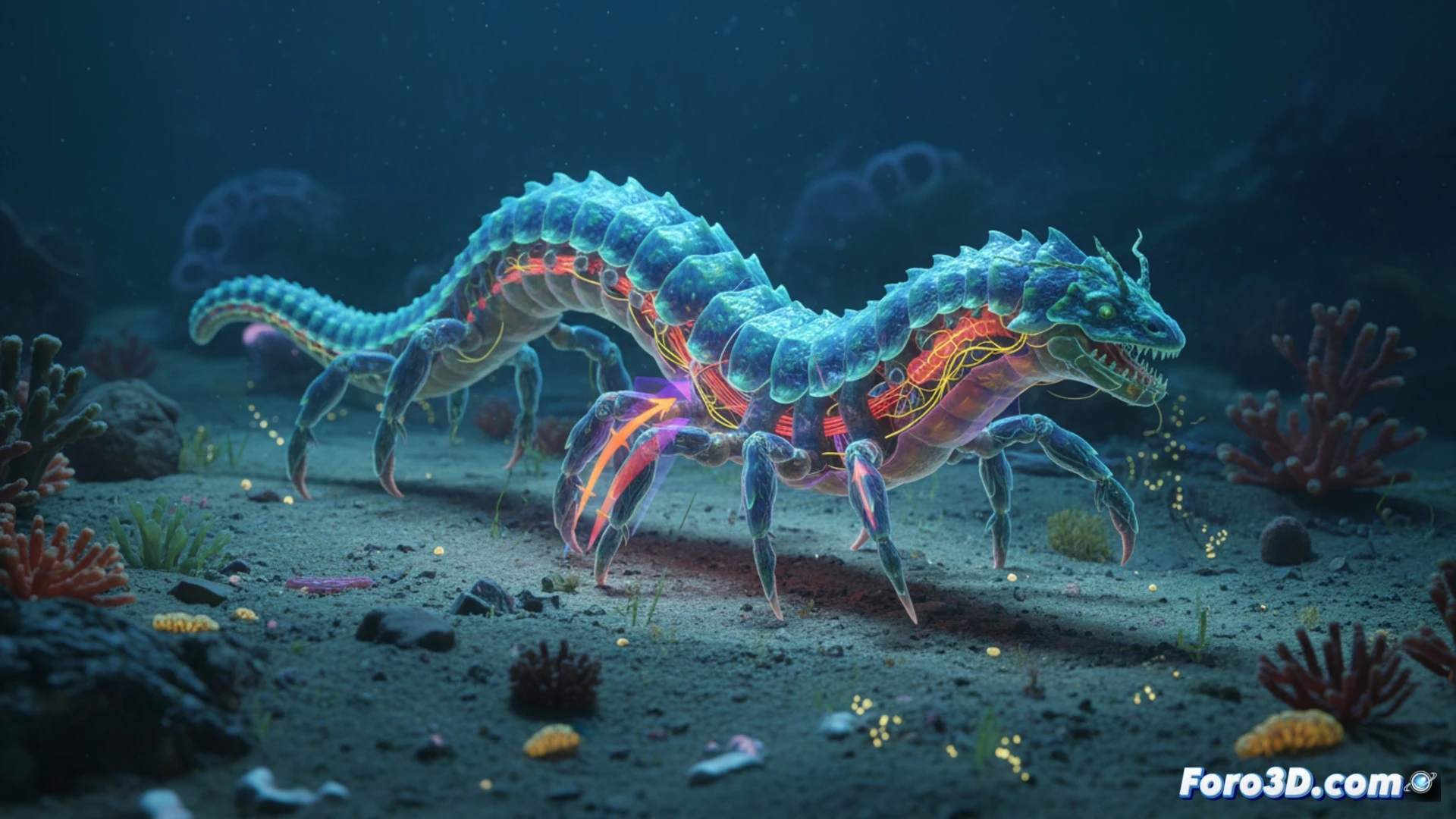
The Dracograllus miguelitus, known as the Dragon Nematode, is a millimeter-sized marine worm that defies the locomotor limitations of its phylum. Unlike typical nematodes, which move through body undulations, this species has developed specialized structures that allow it to walk on the seafloor. For the Scientific Visualization niche, this organism represents a perfect challenge: creating a 3D model that not only displays its external morphology but also reveals the internal mechanisms that make this unique behavior possible. 🐉
Anatomical modeling and locomotion simulation 🦾
The technical proposal consists of developing a 3D animation of the Dragon Nematode in a textured seafloor environment. The model must include a partial transparency layer to visualize the internal anatomy: the hydrostatic muscular system, collagen fibers, and cuticular projections that act as legs. The locomotion simulation requires non-standard rigging, based on inverse kinematics for the locomotor structures and a particle engine to represent interaction with the sediment. A morphological comparison with other marine worms, such as polychaetes, will be added, highlighting differences in the arrangement of muscle bundles. Interactive labels, activated by click or hover, will display scientific data: average length (0.8 to 1.2 mm), locomotion speed (0.5 cm/min), and pressure exerted by the legs on the substrate. The result will be an ideal educational resource for marine biology, accessible from web browsers thanks to WebGL export.
Reflection on scientific visualization tools 🔬
The choice of Blender for modeling and Unity for interactivity responds to the need for balance between anatomical fidelity and real-time performance. However, the greatest challenge is not technical, but conceptual: translating complex biological data, such as fluid mechanics in the nematode's pseudocoelom, into a clear and distortion-free visual representation. This animation not only documents a discovery but also invites us to wonder how many other biomechanical adaptations go unnoticed in marine microfauna. Scientific visualization, when well executed, becomes the bridge between microscopic observation and public understanding of evolution.
How can the biomechanics of the undulatory movement of Dracograllus miguelitus be modeled in an interactive 3D environment to visualize force transfer in its millimeter-scale anatomy
(PS: at Foro3D we know that even manta rays have better social bonds than our polygons)