A knee implant failed prematurely due to loss of fixation with the bone. The investigation revealed that the hydroxyapatite (HA) coating, designed to promote osseointegration, had detached from the metal surface. 3D analysis using scanning electron microscopy and specialized software identified the root cause: an incorrect temperature during the plasma spray process that generated poor interfacial adhesion.
Workflow: from microtomy to finite element simulation 🔬
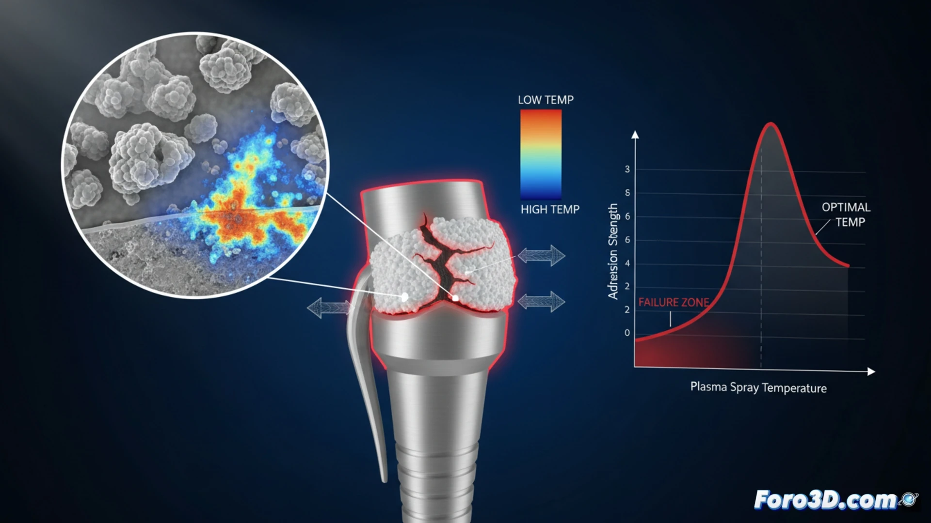
The analysis process began with the acquisition of high-resolution images of the delaminated coating using a scanning electron microscope (SEM) with 3D capabilities (ZEISS ZEN). The volumetric images were segmented in Materialise Mimics to reconstruct the porous geometry of the HA layer, identifying areas of detachment and subsurface cracks. This geometry was exported to Abaqus, where a finite element simulation was performed. The model applied typical physiological gait loads and varied the interfacial friction coefficients, demonstrating that a plasma spray temperature below 40 degrees Celsius drastically reduces adhesion energy, initiating delamination in early loading cycles.
Lessons for quality in orthopedic implants 🦴
This case underscores the need for rigorous control of manufacturing parameters in bioactive coatings. The combination of 3D microscopy and numerical simulation not only explains the failure but also allows establishing optimal process windows for plasma spray. For engineers and surgeons, this methodology becomes an indispensable validation tool, ensuring that the bone-implant bond does not depend on a critical and avoidable factor such as application temperature.
As a researcher, which microstructural factors and fracture mechanisms did you identify as critical in the delamination of the hydroxyapatite coating on the failed knee implant through 3D analysis of its failure surface?
(PS: and if the printed organ doesn't beat, you can always add a little motor... just kidding!)