A recent study in Current Biology revealed that female Aedes aegypti mosquitoes, vectors of diseases like dengue, regulate their blood appetite through specialized cells in their rectum, not in the brain. This finding opens a new avenue for controlling their bite. From scientific 3D visualization, this poses a fascinating challenge: modeling and animating this complex physiological system to understand and communicate the process in an intuitive and precise way.
3D Modeling of Anatomy and Peripheral Neuronal Signaling 🧠
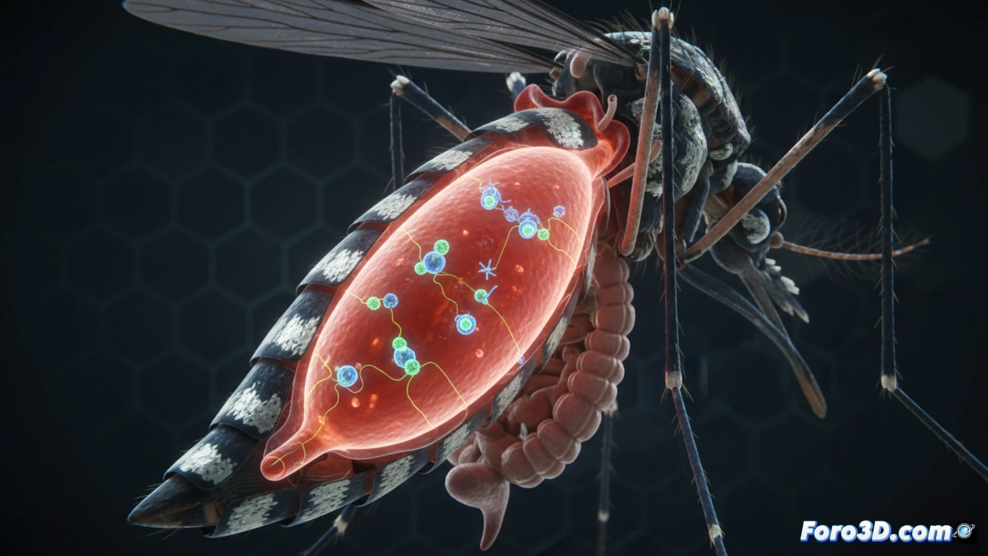
The visualization opportunity lies in creating an interactive 3D anatomical model of the mosquito. The rectal pads could be highlighted, showing the exact location of the key receptors. Then, through procedural animation or simulation, the signal pathway would be visualized: from blood ingestion, neuropeptide release, activation of rectal cells (which act as neurons), and finally the sending of the inhibitory signal to the brain. A slider would allow comparing appetite and satiety states in real time.
From 3D Simulation to Public Health Strategies 🦟
Visualizing this mechanism in 3D is not just academic. An accurate model can be the basis for simulations that test, in silico, receptor-blocking molecules. By visually understanding the spatial interaction, the design of interventions would be accelerated. Thus, scientific visualization becomes a critical bridge between a biological discovery and its potential application to reduce disease transmission.
How would you animate the behavioral patterns described in the study?