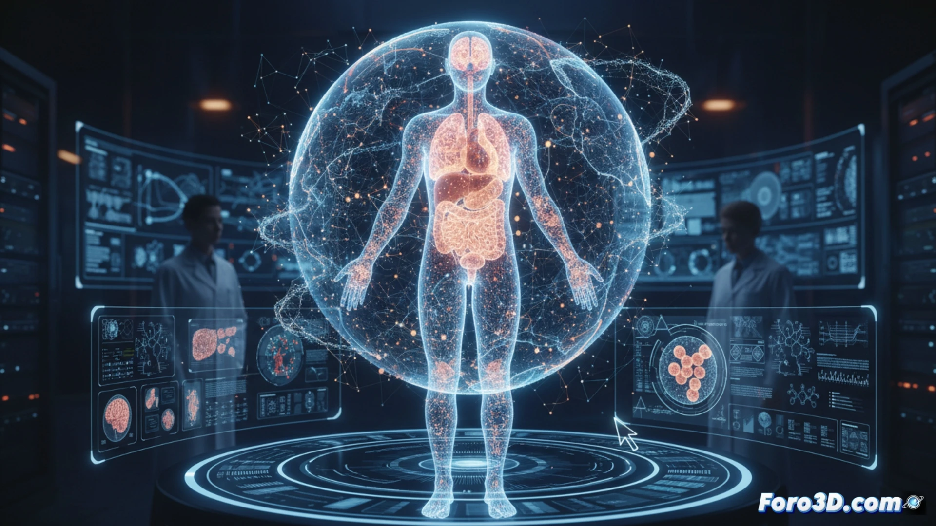
An international scientific consortium has presented a 3D human organs atlas with unprecedented detail. This tool, accessible online, allows interactive exploration of more than 50 organs with nearly cellular resolution. Developed over six years, it promises to revolutionize biomedical research and anatomical education, bridging the gap between radiological images and microscopic tissue studies.
Hierarchical phase-contrast tomography: the key to extreme detail 🔬
The core technology is hierarchical phase-contrast tomography, which uses synchrotron light from the ESRF in Grenoble. This extremely bright source allows non-destructive scanning of entire organs without the need to slice them. The process generates volumetric data with a resolution up to 50 times finer than the thickness of a human hair. The resulting datasets, available for download, are visualized in the browser using interactive tools that allow navigating through tissues like in a digital map, zooming from the organ scale to subcellular details.
A revolution for science and visual education 📚
This atlas is not just a technical achievement; it is a transformative resource. For researchers, it accelerates the study of diseases and comparative anatomy. For physicians, it offers deep structural understanding. For students, it is an unparalleled educational tool. The project envisions extending this technique to entire bodies, marking the beginning of a new era in the scientific visualization of the human body.
How can the scientific visualization of this 3D cellular atlas transform biomedical research and medical education? 🧠
(PS: fluid physics for simulating the ocean is like the sea: unpredictable and you always run out of RAM)