Medicine is advancing toward a future where the first incision is not made in the operating room, but in a digital environment. Virtual twins, exact computational replicas of human organs, are transforming high-risk surgery. These models, created from the patient's medical images, faithfully simulate physiological behavior, allowing medical teams to practice and plan complex interventions beforehand and without risk to the patient.
From scanner to simulator: engineering a digital heart 🔬
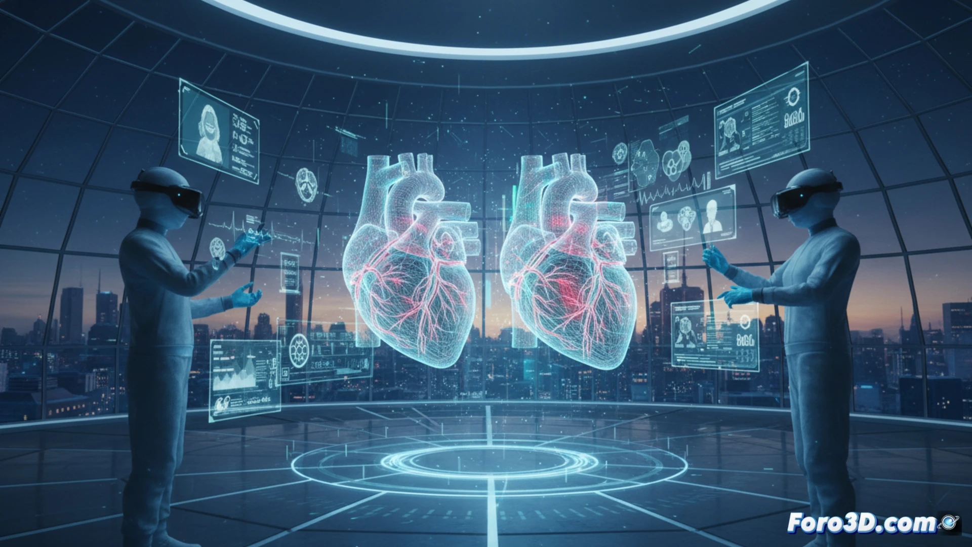
The process begins with patient tomography or MRI data. Using specialized software, a precise 3D geometry of the organ is reconstructed—in this case, the heart. Then, mathematical models are applied that simulate physical properties such as tissue elasticity and fluid dynamics to replicate blood flow. Projects like the Living Heart have perfected this technology, achieving twins that predict how the organ will respond to a suture or a bypass. This allows testing multiple surgical strategies in silico to identify the optimal one, drastically reducing uncertainty on the operating table.
Beyond the operating room: the future of personalized medicine 🚀
Success in thousands of cardiac procedures is just the beginning. This technology promises to extend personalization to all levels of medical care. A digital twin can evolve with the patient, serving as a tool for continuous diagnosis and to test pharmacological treatments virtually. It represents a paradigm shift: from reactive medicine to a predictive and preventive practice, where every clinical decision is backed by an individual-specific simulation, saving more lives with greater precision.
How are digital heart twins revolutionizing surgical planning and reducing risks for patients?
(P.S.: don't forget to update the digital twin, or your real twin will complain)