A team of Swiss researchers has achieved a crucial breakthrough in regenerative medicine by developing auricular cartilage through 3D bioprinting. Using human cells, they have created a tissue that maintains its shape and elastic properties after being implanted in animal models. This work promises to revolutionize ear reconstruction, currently dependent on the extraction of costal cartilage, a painful procedure with less flexible results.
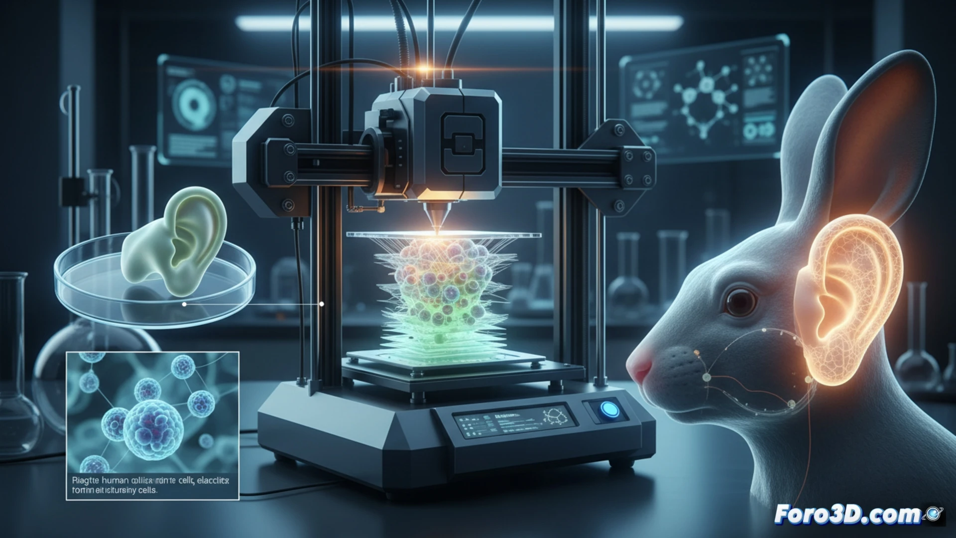
From cell culture to bioprinting: a detailed technical process 🔬
The process begins with the extraction of chondrocytes from small samples of human cartilage. These cells are multiplied in the laboratory and then mixed with a specialized bioink. The mixture is 3D printed adopting the precise shape of an ear. The printed structure is not functional immediately; it must mature in a bioreactor that simulates bodily conditions. There, the cells produce extracellular matrix, developing essential components like type II collagen, key to the tissue's strength. However, the generation of elastin, the protein responsible for flexibility, remains a pending challenge for the team.
The future of personalized biological implants 🚀
This project underscores the potential of biofabrication to create living and personalized implants. The main advantage lies in less invasive reconstructive surgery with more natural results. Resolving the elastin challenge in the next five years could open the door to clinical trials, marking a milestone in the clinical application of tissue engineering and 3D printing in biomedicine.
Could personalized 3D bioprinted auricular cartilage become the standard for ear reconstruction in plastic and reconstructive surgery?
(PS: If you print a heart in 3D, make sure it beats... or at least that it doesn't cause copyright issues.)