The planning of complex cardiac surgeries takes a qualitative leap with the development of a 3D-printed artificial heart. This model, an exact replica of a patient's anatomy created from their medical images, allows surgical teams to practice and simulate the real intervention. This innovation in 3D biomedicine offers risk-free training, improves procedure precision, and personalizes treatment, translating into greater safety and better clinical outcomes.
From scanner to operating room: the technical workflow 🧠
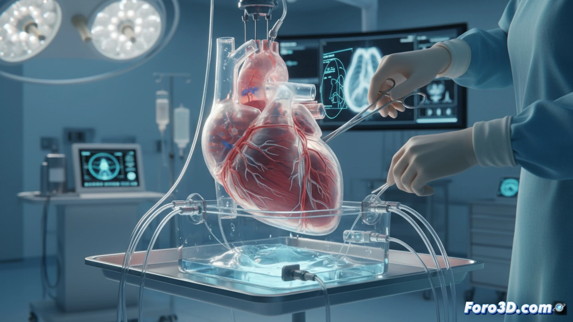
The process begins with the acquisition of high-resolution images of the patient, using computed tomography or magnetic resonance imaging. This data is processed with specialized software to segment and isolate cardiac structures, generating a precise 3D digital model. This file is prepared for printing, selecting materials that simulate the consistency of real tissues. The resulting physical twin faithfully replicates malformations, valves, and blood vessels, providing a tangible object for surgeons to evaluate approaches, test instruments, and anticipate complications in a controlled environment.
Beyond the model: impact on the future of surgery 🔮
This technology goes beyond the mere creation of a visual replica. By offering a tactile and realistic simulation, it reduces uncertainty in the operating room, shortens operation times, and minimizes risks to the patient. It represents a crucial step toward fully personalized surgery, where each intervention is designed and optimized on the patient's unique organ. Its adoption promises to establish a new standard of precision in cardiovascular medicine.
What segmentation software do you recommend for this medical data?