Cardiac micro-cavitation is a biomechanical phenomenon that occurs when the intracavitary pressure of the heart drops below the vapor pressure of blood, generating microscopic bubbles. This process, linked to high-intensity ultrasound procedures or the operation of implantable devices, can cause everything from silent embolisms to severe tissue damage. Understanding its dynamics is critical for clinical safety.
Biomechanical modeling of bubble dynamics 💧
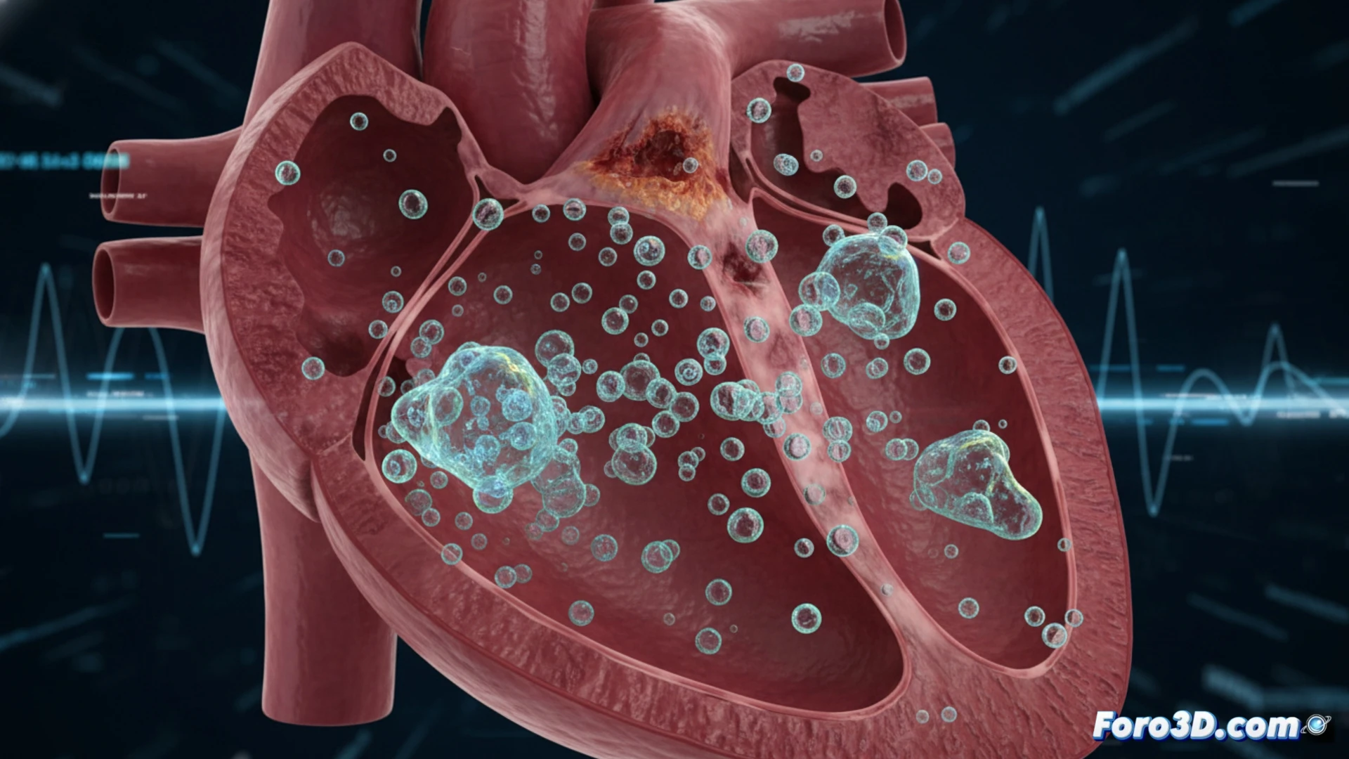
3D modeling allows reproducing the formation, growth, and collapse of these bubbles in cardiac cavities through finite element simulations. Software such as COMSOL Multiphysics or Ansys Fluent integrates Navier-Stokes equations with Rayleigh-Plesset cavitation models, coupled with the real geometry of atria and ventricles obtained from tomographies. Anatomical visualizations show how bubbles concentrate in low-pressure areas, such as the ventricular apex or valves, facilitating the prediction of risk points in interventions like catheter ablation or left atrial appendage closure.
Towards a safer 3D cardiology 🫀
The integration of these simulations into surgical planning and cardiologist training allows anticipating adverse events without exposing real patients. Additionally, educational animations generated from these models help explain to patients why certain procedures require continuous ultrasound monitoring. Micro-cavitation ceases to be an abstract concept and becomes a controllable variable within the ecosystem of three-dimensional digital medicine.
How can 3D simulation of cardiac micro-cavitation help predict the risk of tissue damage during procedures with ventricular assist devices?
(PS: If you 3D print a heart, make sure it beats... or at least that it doesn't cause copyright issues.)