When a heart valve fails, blood flow becomes turbulent and dangerous. 3D Biomedicine offers a radical solution: transforming CT scans into precise three-dimensional models. These digital twins allow surgeons to observe the defective anatomy from any angle, accurately measuring the degree of stenosis or insufficiency before touching the scalpel.
Segmentation and hemodynamic modeling 🩺
The process begins with the segmentation of DICOM images obtained from CT or MRI scans. Using specialized software, the valve tissue is isolated from the rest of the heart, creating a polygonal mesh that replicates every fold and calcification. This model is 3D printed with flexible materials that simulate the elasticity of real tissue. CFD (computational fluid dynamics) simulation adds the hemodynamic variable: pressure and blood flow data are injected to visualize areas of turbulence and mechanical stress, quantifying the exact severity of the failure.
Tactile validation before the operating room 🖐️
Holding a 3D-printed valve changes surgical planning. The medical team can rehearse the repair or deployment of a transcatheter prosthesis on the physical model, identifying risks of paravalvular leak or structural rupture. This practice reduces ischemia time in actual surgery and improves procedure precision, turning an abstract diagnosis into a tangible, life-saving experience.
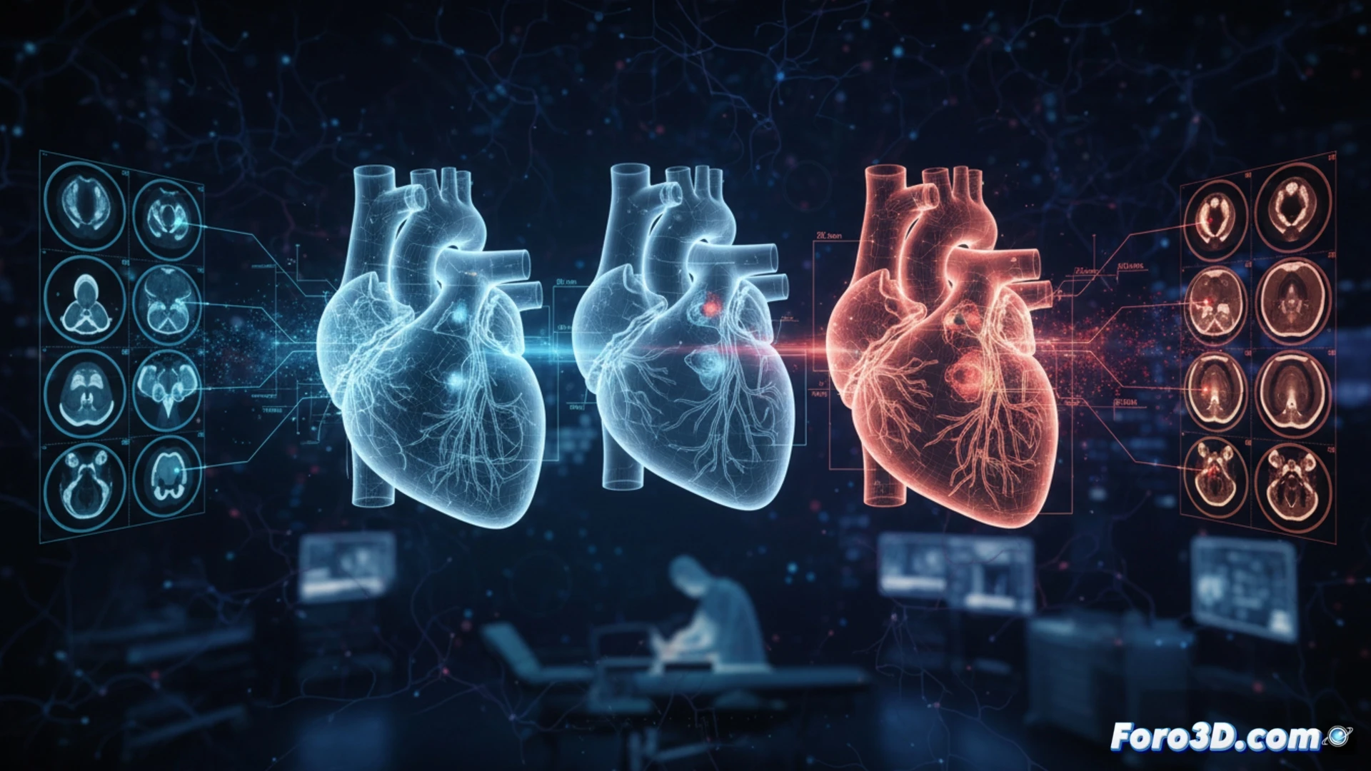
How a digital twin of a heart valve is designed to accurately predict blood turbulence before a surgical intervention in 3D Biomedicine
(PS: If you 3D print a heart, make sure it beats... or at least doesn't cause copyright issues.)