The recent news about a critical failure in a tissue bank has brought the fragility of biological preservation processes to the center of the debate. When we talk about tissues intended for grafts and transplants, any error in the cold chain or structural manipulation can result in the total loss of the material. This is where 3D technology ceases to be a laboratory luxury and becomes an indispensable clinical safety tool.
Cryopreservation simulation and 3D-printed scaffolds 🧊
3D modeling allows recreating digital twins of organs and tissues to simulate cryopreservation processes before applying them to real samples. Through finite element analysis, it is possible to predict the formation of ice crystals or the uneven distribution of cryoprotectants, identifying failure points without risking donated tissue. In parallel, 3D printing of biocompatible scaffolds offers a synthetic backup alternative. These scaffolds, made from hydrogels and polymers, can be stored as emergency stock, reducing exclusive dependence on cadaveric tissues and minimizing the impact of a logistical failure.
Inventory digitization as insurance against human error 📋
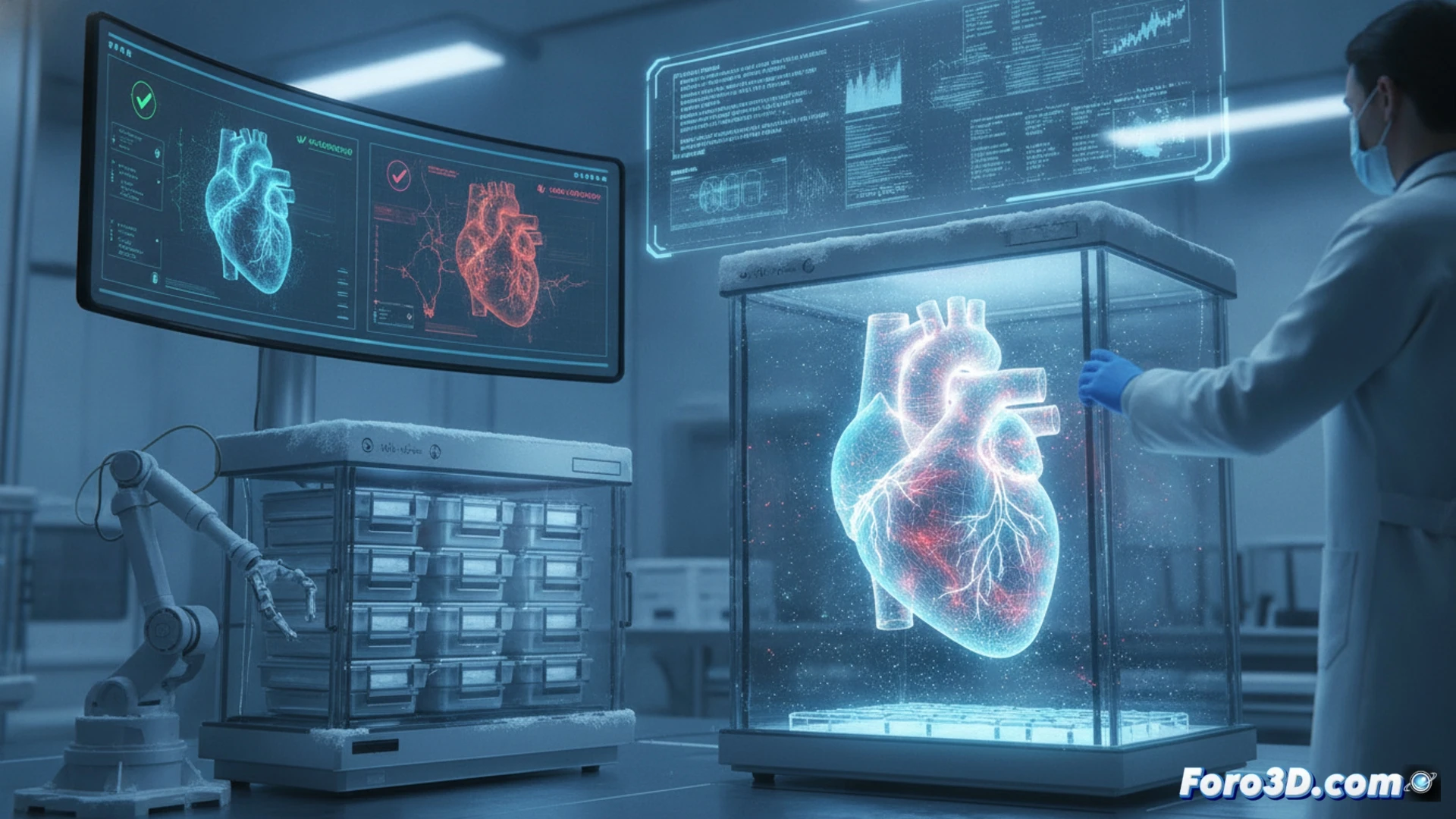
Beyond biology, failure often has an administrative or handling origin. Complete digitization of a tissue bank using 3D scanners and volumetric tracking systems allows creating a virtual inventory where each sample has a unique geometric footprint. This eliminates identification confusion and enables remote audits. A digital twin of the storage container can alert about thermal deviations in real time, transforming passive management into a proactive system that anticipates failure before the tissue degrades.
What role can 3D digital twins play in the early detection of failures in the cold chain of tissue banks?
(PS: If you 3D print a heart, make sure it beats... or at least that it doesn't cause copyright issues.)