A recent clinical case has brought a critical problem in traumatology to the forefront: the fixation failure of an orthopedic implant. When an external fixator or prosthesis does not anchor correctly to the bone, the result can be a failed recovery and the need for a second surgery. This article explores how 3D printing of patient-specific biomodels allows surgeons to identify these weak points before entering the operating room, transforming surgical planning into a predictive process.
Preoperative biomechanical analysis with anatomical models 🦴
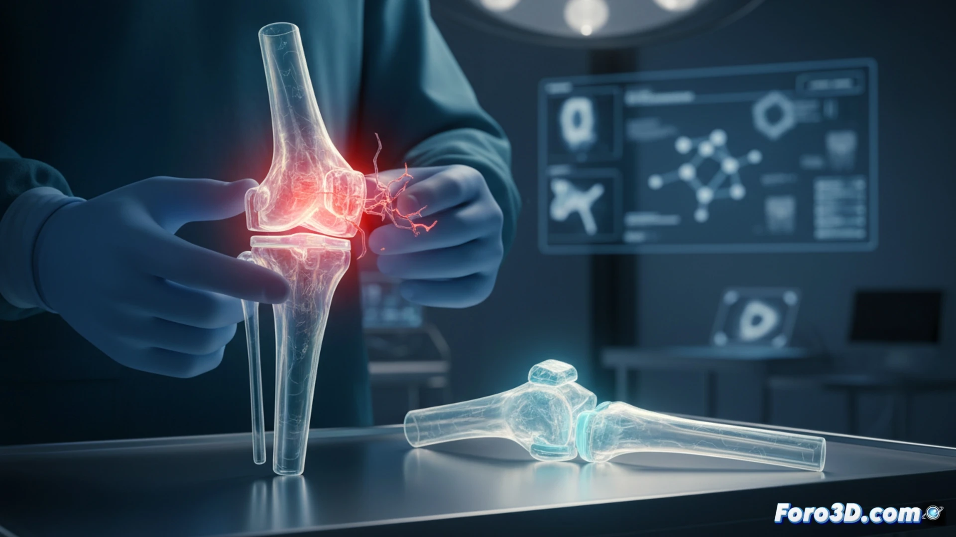
3D printing technology allows for the fabrication of exact replicas of the patient's bone anatomy using computed tomography (CT) data. These models, printed in materials that simulate the density of cortical and trabecular bone, enable mechanical stress testing. In the case of a fixation failure, the surgical team can mount the actual hardware (screws, plates, or fixators) onto the printed model. By applying controlled forces, micro-movements or high-tension points that could cause postoperative loosening are detected. This physical simulation allows for adjusting the screw insertion angle or changing the plate position without risk to the patient.
Surgical prevention through tangible simulation 🔧
The main lesson from fixation failure is that reliance on 2D digital planning is no longer sufficient. 3D printing offers tangible validation that no software can match. By holding a printed model, the surgeon can visualize the bone-implant contact and verify rotational stability. Incorporating this practice routinely not only reduces reoperation rates but also shortens surgical time by having a proven fixation strategy. The failure thus becomes a lesson for standardizing the use of biomodels in orthopedics.
Can 3D printing of custom implants with gradual porous structures eliminate the risk of failure due to micromotion and bone necrosis in patients with low bone mineral density?
(PS: and if the printed organ doesn't beat, you can always add a little motor... just kidding!)